
In this topic, we explore the structure and function of the nervous system. Because the nervous system is responsible for tasks that help maintain life (homeostasis), there is a lot of detail. Consider adding any terms you are not sure of to your personal terminology and vocabulary reference guide. Don’t be afraid if there are a few, because there is a lot of science involved in the nervous system. The payback is that it’s all super cool!
The nervous system is responsible for a vast number of functions in the body, all with the same aim; keeping controlled conditions within limits that maintain life (homeostasis). However, the nervous system does not do this alone. This is an example of when two or more systems work together to maintain a safe functioning working environment of the human body.
Think of the nervous system as the 'Godfather'. It is the receiver of all messages from the other ten body systems and is quite literally the brains of the operation (pardon the pun). The nervous system (the brain is the other component and discussed later):
- receives these messages
- translates them as to what, why, where and when attention is needed and how it will perform this action in the body
- communicates this to fellow systems and lets them know what part they place in this.
This is teamwork!
Understanding the nervous system and its components (of which there are a few) will help you understand how muscles work in terms of exercise and when working out and cooling down. This is important because a solid grasp on this information will equip you to be confident and knowledgeable whilst providing a safe and valuable experience for your clients.
In this topic, we focus on the structure and function of the components within the human nervous system. You will learn:
- the nervous system and homeostasis
- the functions and divisions of the nervous system
- the structure of the nervous system
- the anatomy of a neuron
- the structure and function of the synapse and nerve communication
- how electrical impulses are generated
- receptors involved in sport.
Terminology and vocabulary reference guide
As an allied health professional, you need to be familiar with terms associated with basic exercise principles and use the terms correctly (and confidently) with clients, your colleagues, and other allied health professionals. You will be introduced to many terms and definitions. Add any unfamiliar terms to your own vocabulary reference guide.
Activities
There are activities throughout the topic and an end of topic automated quiz. These are not part of your assessment but will provide practical experience that will help you in your work and help you prepare for your formal assessment.
Ever wondered how the nervous system did its job? Let’s start with an overview:
The nervous system carries out numerous functions within the human body. It enables us to produce speech, sense several smells, remembers past information, memories, and new ones, provide signals which control body posture and movements, and also regulates bodily organs, just to name a few!
These functions are subdivided into three basic functions:
- Sensory function of the nervous system
- Integrative function of the nervous system
- Motor function of the nervous system.
Note: these functions are fully classified as Sensory: Input, Integrative: process and Motor: Output
Sensory function of the nervous system
The sensory receptors detect stimuli. They detect information such as an increase in body temperature or external stimuli such as a cold breeze. This sensory information which is received, for example by the hairs on our skin, is then carried to the brain and spinal cord for processing through the spinal and cranial nerves.
Integrative function of the nervous system
This is when the nervous system performs its show-stopping performance in analysing the information received and then making a decision as to how and when to respond, and what in the body to recruit to respond appropriately. This decision-based process is known as integration.
Motor function of the nervous system
This is where we see the whole performance coming together. The nervous system flaunts its talents and sends messages (also known as motor responses) which activate organs such as the muscles to move and/or the glands to secrete their substances.
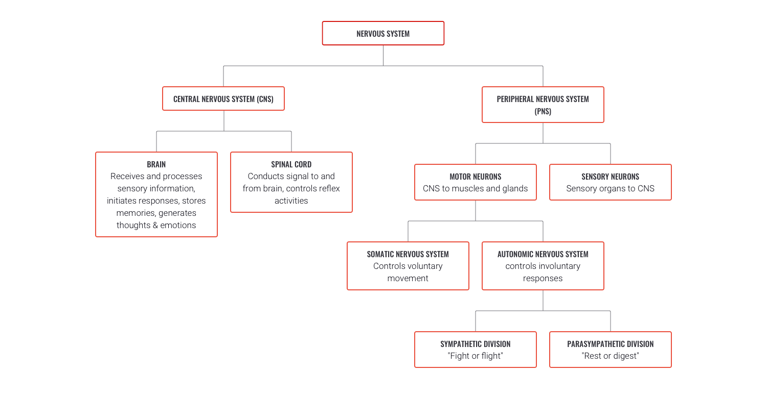
While there are many components making up the entire nervous system, the three general classifications for components include:
- The brain: The brain is the supercomputer of the body. It processes billions of pieces of information and decides how and what we should focus on, respond to, or ignore.
- The spinal cord: The spinal cord is the superhighway for communication. Carries messages between the peripheral nerves and the brain. This also stores vital reflex commands for flight or fight responses.
- The neurons (nerves): Neurons are the pathways of communication between the body parts and the spinal cord. They also carry impulses up the spinal cord and to and from the different parts of the brain.
Let's take a look at each component in closer detail, starting with the brain.
The brain
The brain is made up of about 85 billion neurons! They equip the brain with the communication tools it needs to perform its function (there’s our friend 'structure and function' again!) The adult brain is anatomically subdivided into four major parts:
- Cerebrum
- Diencephalon
- Cerebellum
- Brain stem.
It is important to have a comfortable understanding as to their general functions, so let's explore each part.
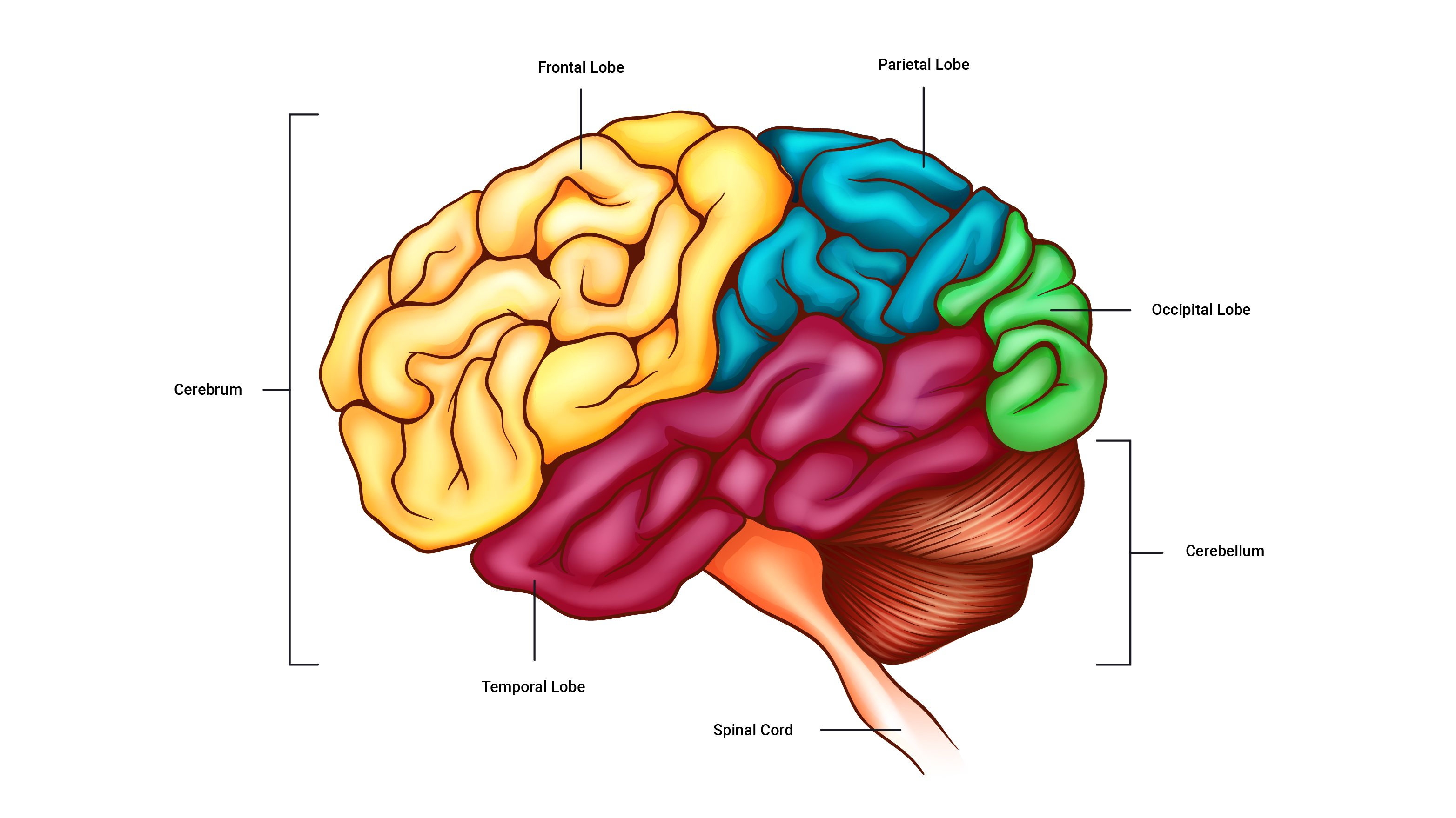
The cerebrum
The cerebrum is the largest part of the brain. It is comprised of four lobes responsible for different processes and actions. For example, thinking, movement control, sensory perception, and sight. Let's have a closer look at the specific function of each of the four lobes.
Frontal lobe
The frontal lobe is at the front of the brain and responds to impulse pertaining to conscious thought, motor control, speech, memory, behaviour.
Parietal lobe
The parietal lobe sits behind the frontal lobe (posterior to the frontal lobe). It is responsible for responding to impulses related to the senses of feeling, touch, pressure, pain and language comprehension.
Occipital lobe
Occipital, referring to the back of the head identifies the location of this lobe of the cerebrum. This region plays an important role in responding to impulses regarding vision by helping us process shapes, colours, movement. It also supports us to interpret and draw conclusions about what we see.
temporal lobe
The temporal lobe is located in the temple region. The temporal lobe is responsible for interpreting impulses relating to hearing, learning, feelings/emotions, vision and memory.
The diencephalon
The diencephalon is where we find the hypothalamus, as well as other parts which are responsible for controlling the body's internal environment, such as temperature control, regulation of heart rate and breathing, hunger, sleep and wake cycles and more.
The cerebellum
The cerebellum sits behind the brain stem and co-ordinates movements and helps maintain body awareness.
The brain stem
The brain stem continues from the spinal cord and essentially connects the spinal cord to the brain. The brain stem has regulatory functions involved in breathing and heart rate.
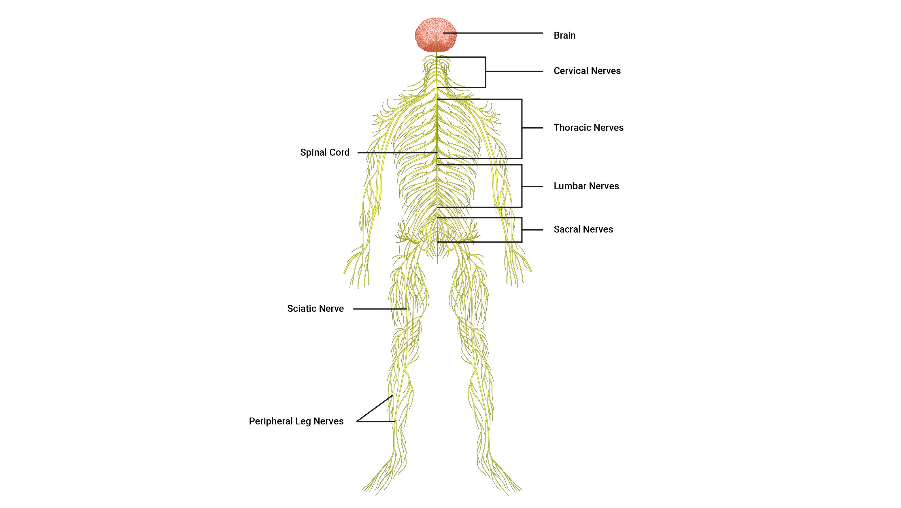
The spinal cord
The spinal cord is a long, thin, incredibly fragile tubular structure encased within the spinal column and surrounded by cerebrospinal fluid. It is composed of nervous tissue and tracts of nerve fibres that allow two-way conduction of nerve impulses, both sensory and motor (input and output) this is important to remember as we progress through this topic.
The third classification for the components that make up the nervous system is the neurons (nerves). Nerve anatomy is a large and complex area so we will look at this next.

Neurons
Neurons are the individual pathways that carry electrical impulses around the human body. Nerves are bundles of neurons all heading in the same direction with neurons branching off them at intervals to get to their intended destinations.
Neurons are one of two types of nervous tissue cells: neurons and neuroglia. In this topic, we focus on neurons. Neurons are responsible for producing complex networks and connections between the brain and the spinal cord and between the rest of the body and the brain and spinal cord.
These lines of communication are composed of highly specialised cells capable of transmitting highly complex information at great speeds. To learn more about how they achieve this, it is important to note there are three main types of neurons in the body which tag team each other in the journey of signal transmission. These are the:
- sensory neuron
- interneuron
- motor neuron
Study the following illustration which shows the three types of neurons.

Fun fact!Our nervous system is the smallest of all 11 body systems and the most complicated with a hugely important role. With this in mind, surprisingly, it accounts for approximately only 2kg which is approximately 3% of our total body weight!
Let's look at the three types/classifications of neurons.
Functional classification of neurons
Sensory neurons
Sensory neurons do as it says on the tin, they 'sense' information and provide the link between sensory receptors and the spinal cord. They transport electrical impulses to the spinal cord where the impulses are passed to interneurons.
Interneurons (Inter~ between)
Interneurons are found only within the spinal cord and brain (CNS) and are situated between sensory and motor neurons. They process incoming sensory information received from sensory neurons and then activate the related motor neuron. They carry impulses up to the brain for processing and decision making. Ninety per cent (90%) of all neurons in the body are interneurons.
Motor neurons
Motor neurons carry impulses that exit the spinal cord away from the central nervous system and are directed towards the effectors of the body, such as muscles, organs, and glands to cause action.
Anatomy of a neuron
In reality, all neuron types look slightly different but contain the same key components. The picture below shows a generalised version of a neuron.
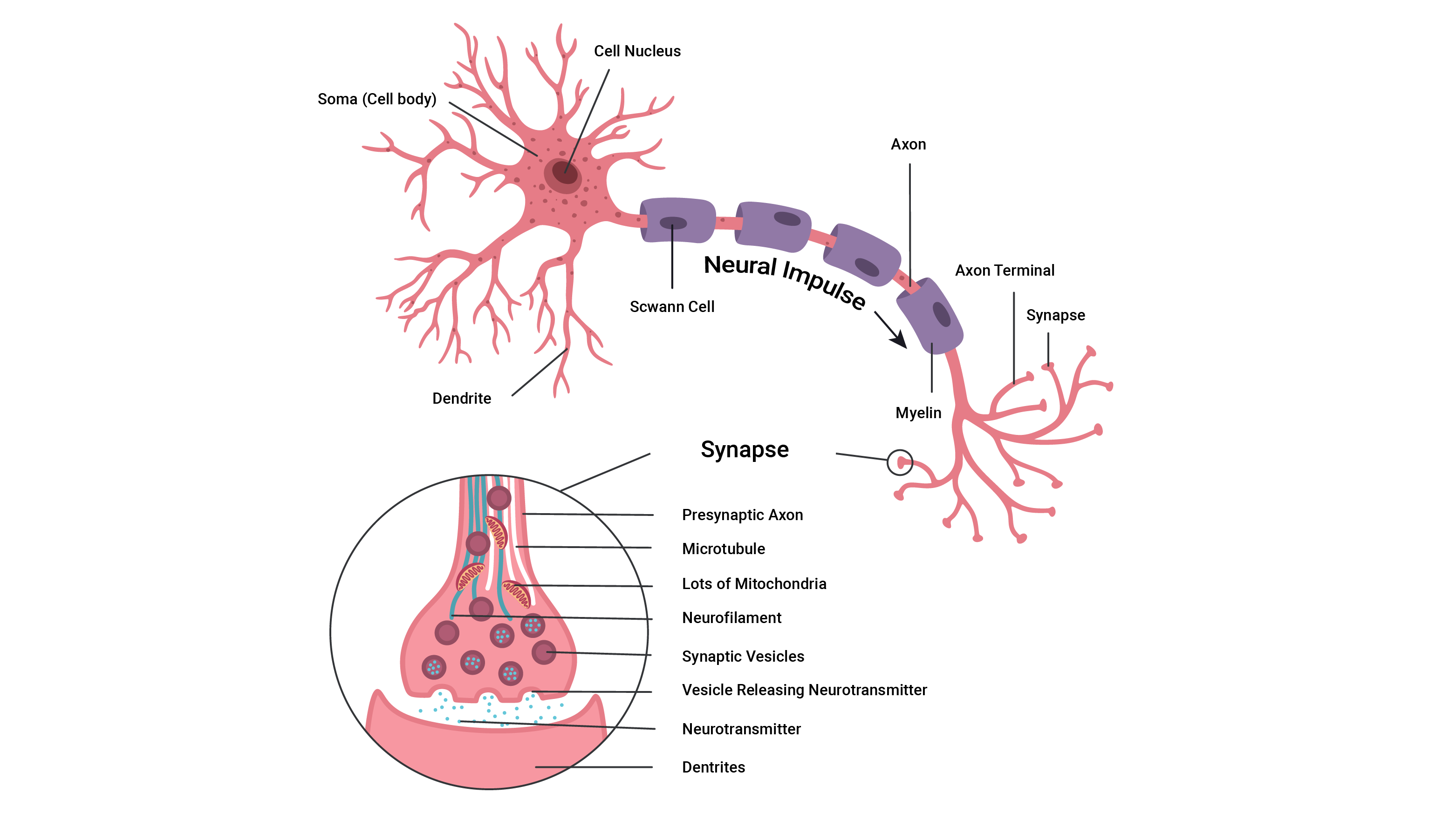
Soma (cell body) and nucleus
- The control centre of the neuron is its cell body (also called the soma).
- It has a centrally located nucleus which acts as the brain of the neuron.
- The cell body usually gives rise to a few thick processes (sticky out bits) that branch into a vast number of nerve fibres known as dendrites.
Dendrites
- Dendrits~ little trees. They do look like little trees and their branches. They house numerous sensory receptors for neurotransmitters and are the primary sites for receiving signals from other neurons.
- The more dendrites a neuron has, the more information it can receive from other cells to incorporate into its decision making.
Myeline sheaths and Schwann cells
- All axons of the PNS (peripheral nervous system) have a sheath of Schwann cells around them. Each Schwann cell spirals repeatedly around the axon, putting down as many as 100 layers.
- These layers form the myelin sheath. Think of the myelin sheath as similar to plastic around an electric cord. The myelin helps fully envelope the axon of the neuron (the highway of electrical transmission pathway) to ensure there are no 'leaks' in information and to help speed the transmission of signals.
- Myelinated nerve fibres conduct impulses faster than unmyelinated nerve fibres.
Axon
- The axon is cylindrical and relatively un-branched for most of its length. Its function is to allow rapid transmission of nerve impulses from the cell body.
- Axons may range from a few millimetres to more than a metre in length.
- An axon will typically connect with another muscle fibre or gland cell via its 'axon terminals'.
Axon terminals (synaptic knobs/end bulbs)
- At the distal end, axons usually break into a complex network of fine branches. Each branch ends in a little swelling called a 'synaptic knob/bulb' (presynaptic terminal that comes before the synapse; this will make more sense in the following subtopic).
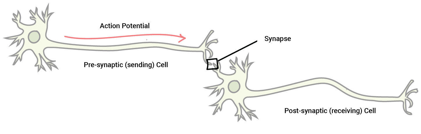
- Each knob forms a junction with a muscle cell, gland cell, or another neuron. The point where communication occurs between two neurons or a neuron and an effector cell is called a Synapse.
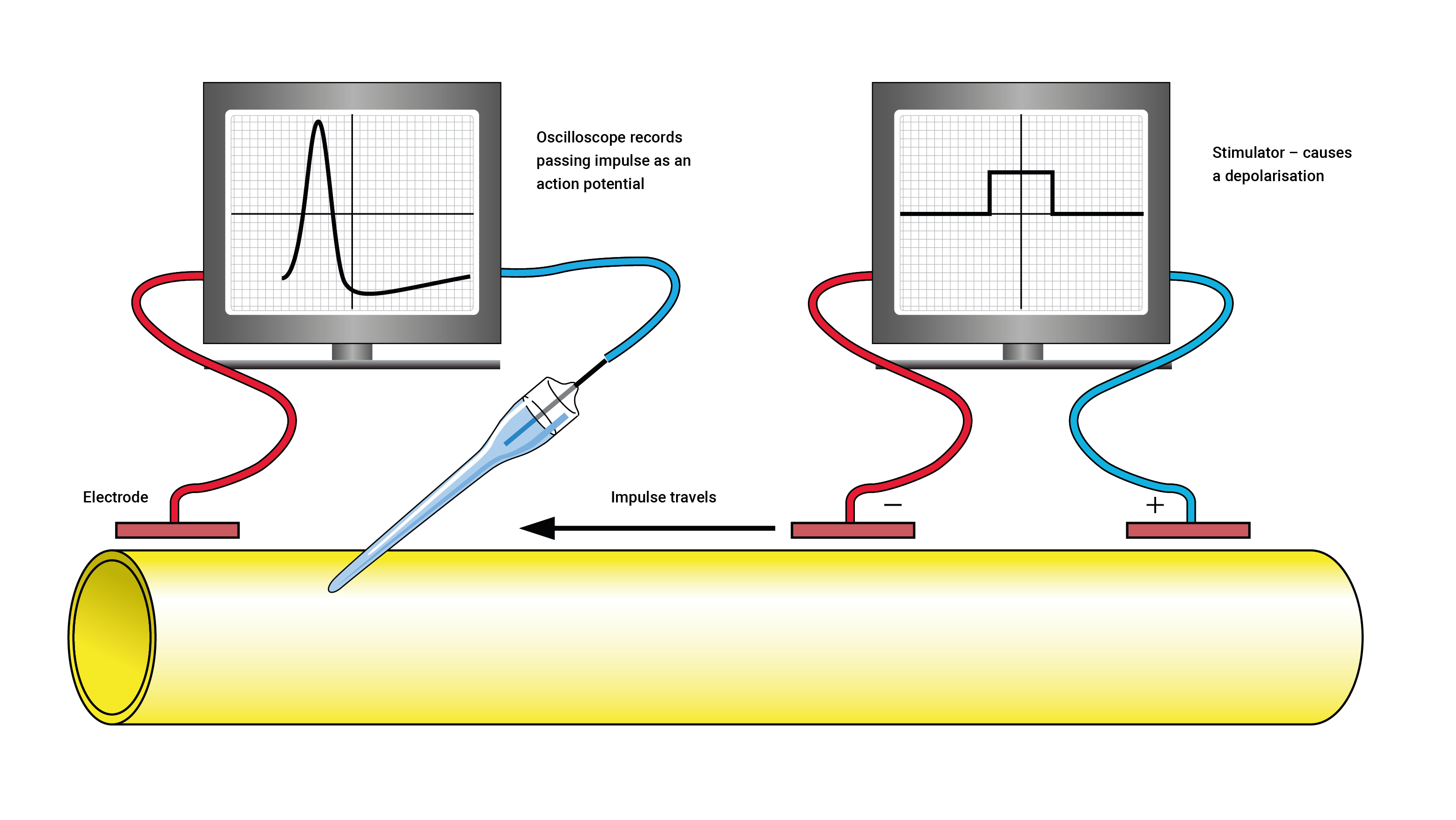
The currency of the nervous system is electrical impulses. These are also known as Action Potentials (AP). It is important to keep in mind (and this will make sense shortly) that these electrical impulses, being electricity, have a voltage.
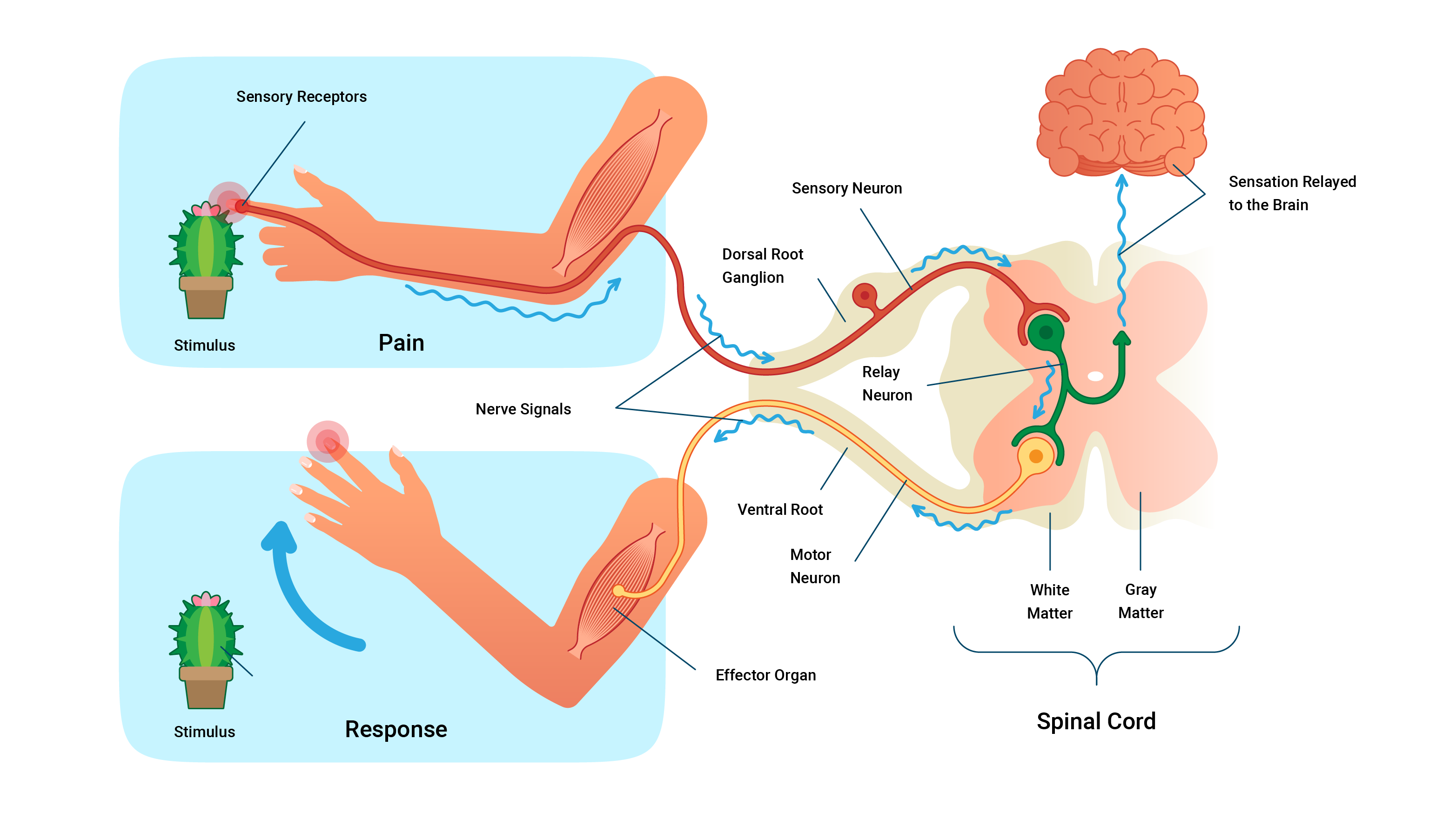
Almost every nerve communication occurs in the same three-step process.
- Sensory receptors notice a change in the internal, or external, environment and send impulses via sensory neurons to the central nervous system (brain and spinal cord) for processing.
- Interneurons in the brain or spinal cord (CNS) process these impulses and decide to act, store the information, or discard it.
- If action is warranted, the CNS sends impulses down motor neurons to the body's effectors to cause action (for example, to the muscles, glands, organs).
The Synapse
A synapse is the site of communication between a neuron and another neuron or a neuron and an effector cell.
These meeting points allow impulses to be passed between the different neurons. The transmission of these electrical impulses contains a few steps and is somewhat complex at first but we will break each of the steps involved.
Physiology of the synapse
The essential components of a synapse are outlined below:
- The pre-synaptic terminal: This is the synaptic end bulb (or knob) of the first neuron. The term 'pre-synaptic' describes its location as coming before the synapse. It contains neurotransmitters packaged into 'vesicles'. Neurotransmitters are chemical couriers that are vital in generating new impulses on the other side of the synaptic cleft.
- The synaptic cleft: A small gap that separates the pre and postsynaptic terminals (that is, the connecting parts of two neurons).
- Impulses can't 'jump' the synaptic cleft, instead, they send neurotransmitters across.
- The postsynaptic membrane: The dendrite of the next neuron, post~ implying 'after'. When neurotransmitters bind to receptors on the postsynaptic membrane, ion channels open allowing positively charged ions to enter, if enough arrive a new impulse is formed and the message is passed on.

How nerves fire
The steps in this process are:
- Electrical impulse (action potential) flows down the body of the presynaptic neuron.
- This positive charge (voltage) encourages ion channels on the membrane of the synaptic end bulb to open and let in calcium Ca2+.
- The influx of positive Ca2+ ions triggers the packages (vesicles) of neurotransmitters to move from the middle of the bulb to the outside and spill their contents into the synaptic gap.
- The neurotransmitters then bind to receptors on the postsynaptic terminal (bulb) and trigger the receptors to open and let in the Na+ ions which then, in turn, continue the positive charge from one neuron to the next. And so the cycle continues!
Note: These steps summarise the communication process that occurs along a neuron and reaches the synaptic gap/cleft. The process that occurs next depends on the neighbouring cell (postsynaptic cell) which could either be another neighbouring neuron or an effector cell such as a muscle cell or a gland. These steps will be discussed in the relevant topics which you will meet later.
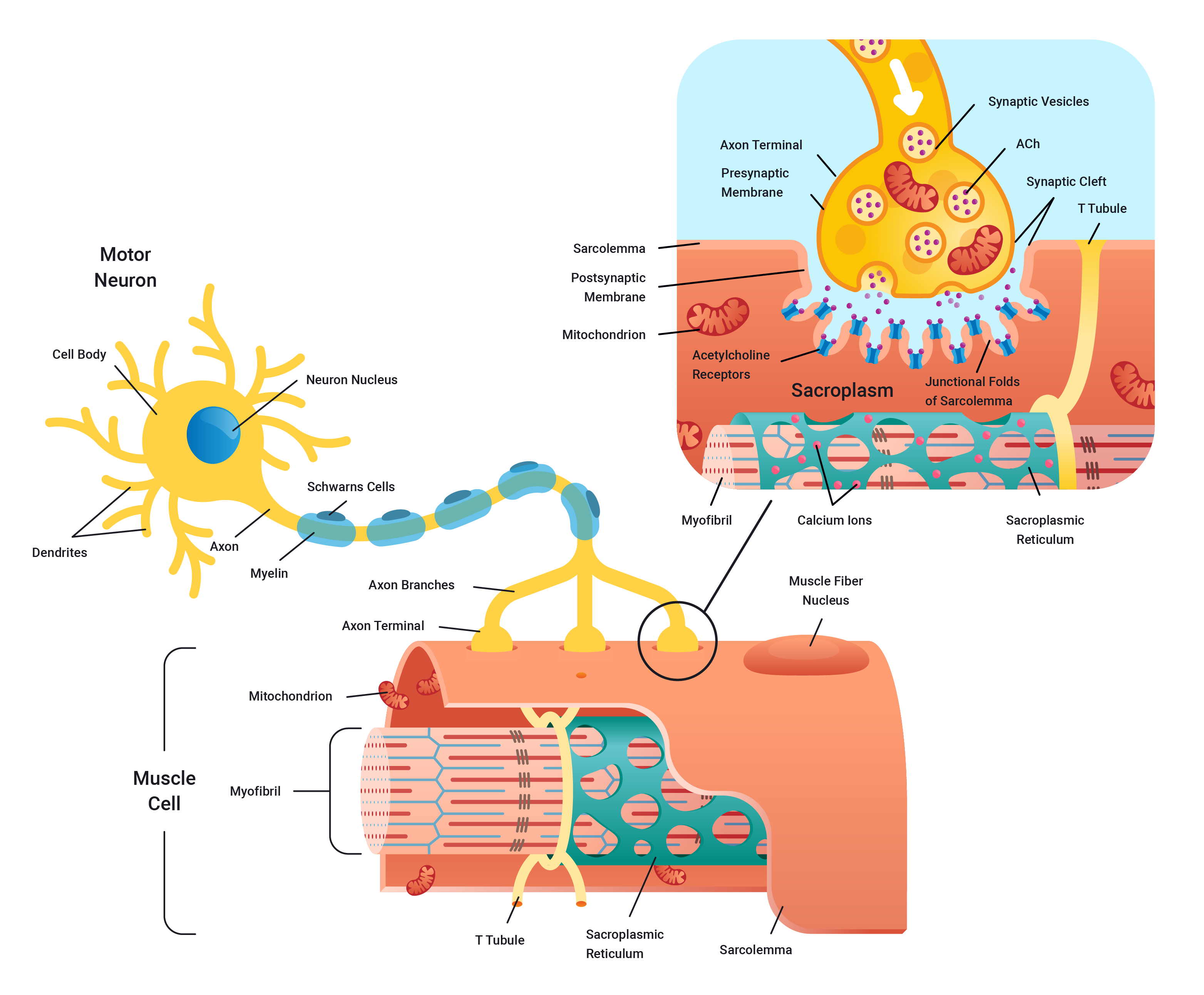
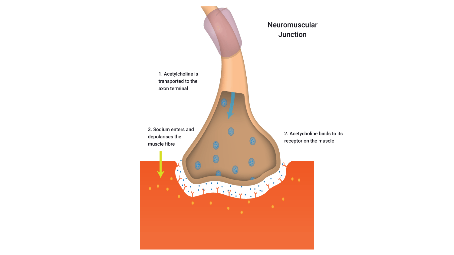
Neuromuscular junction
Watch the following video which shows a concise rundown of the process of communication at the synapse. It is a little longer than other videos and touches on aspects in more detail that you are expected to learn. However, it does offer a great overview which will assist in cementing the concepts you have learnt so far about nerve anatomy and their structure and function.
Nerves conduct electrical impulses, but where does the electricity come from? The electricity needed to send impulses is derived from electrolytes. These are positively charged ions within the body such as sodium, potassium, calcium. Neurons are bathed in an extracellular fluid which is rich in electrically charged ions. The inside of neurons also contains positively charged ions.
Resting membrane potential
The average resting membrane potential (charge) of a neuron is -70mv. As the name implies, 'resting' means it is currently inactive in a sense, it's resting. In order for it to become electrically stimulated (excited) it needs to become more positive (or less negative) which occurs via a process known as depolarisation.
Depolarisation
Depolarisation occurs when a nerve increases its positive charge. This occurs as positively charged ions (for example, sodium Na+/ calcium Ca2+) enter the nerve cell. A nerve must depolarise if it is to fire.
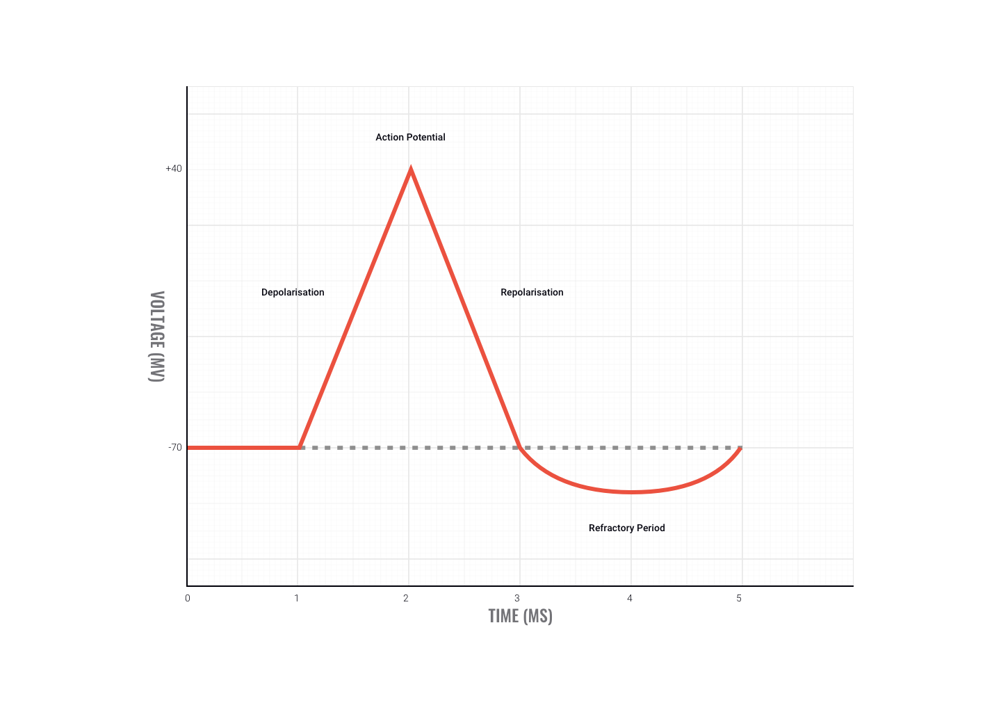
Threshold
In order for a neuron to fire, the depolarisation must be enough to reach the neuron's threshold, i.e. raise the neuron's resting membrane potential above -55mV.
Repolarisation
Repolarisation refers to the stage during an action potential where the neurons positive charge returns towards its resting potential, positive ions flow out of the neuron and as a result, its in charge becomes more negative ( less positive), returning towards -70mv.
All or nothing principle
There is no such thing as a big or small action potential, all action potentials are the same, either the membrane reaches potential and the neuron fires or it doesn't, all or nothing.
Watch the following video which gives a thorough rundown of the before, during, and after action potential generation. Again, it is a little long and has more detail than required at this point. However, sometimes it pays to dig a little deeper to understand the surface knowledge.
The motor divisions of the nervous system include the:
- somatic division
- autonomic division
- parasympathetic division
- sympathetic division.
Take a look at the picture below to see how they align with the central and peripheral nervous system divisions.
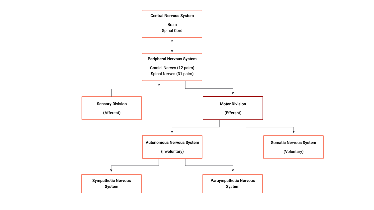
The motor division
Once a decision has been made by interneurons, motor neurons take and transmit the impulses to the effectors of the body, such as the muscles, organs and glands.
The motor division of the PNS (peripheral nervous system) is divided into:
- the somatic nervous system (voluntary)
- the automatic nervous system (involuntary)
These differ from each other by the different types of effectors they innervate (supply).
The somatic division
The somatic division includes nerve pathways of the PNS that lead to skeletal muscle. It creates voluntary movement.
The autonomic nervous system
The autonomic nervous system controls the body's involuntary internal functions, including but not limited to:
- heart rate
- blood pressure
- blood distribution
- respiration
- digestion
- production of saliva, sweat and tears
- urination.
How lucky are we that the autonomic system takes full control of these aspects of human physiology? Imagine if we had to consciously think about breathing in and out, and our heart beating, for example. That would be a nightmare and we would end up in serious problems! Thankfully this is taken care of for us by our new friend, the autonomic nervous system.
The automatic division can be split into two further divisions:
- The sympathetic division
- The parasympathetic division.
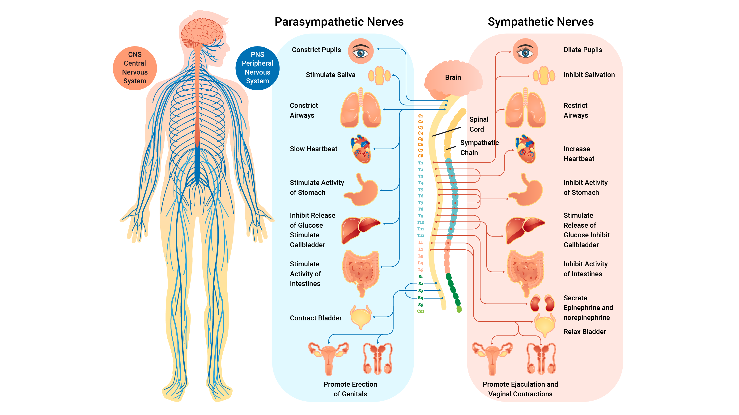
The sympathetic division
The sympathetic division prepares the body for expending energy. It is used in emergencies or stressful situations and also known as the 'Fight, Flight, Fright' response. For example, flinching when someone shouts, when a ball flies towards you, breathing fast when nervous and sweating in a job interview.
The parasympathetic division
The parasympathetic division is the body’s housekeeping system. It has a major role in digestion, producing urine, glandular secretion, and conserving energy. It is most active under ordinary restful conditions and also known as the 'Rest and Digest' system. For example, digesting food, sleep and wake cycles, and regulating breathing and heart rate.

Sensory receptors play vital roles in sport and exercise. They enable us to know where our body parts are in space, help our body determine oxygen requirements during aerobic exercise, and keep us safe by helping to avoid soft tissue injuries. Let's focus on four key sensory receptors including:
Chemoreceptors
These receptors monitor and respond to changes in the chemical environment within the muscle (for example, O2 and CO2 levels, acidity).
They provide the central nervous system (CNS) with information on the metabolic rate of muscular activity. An example of this is reporting increasing oxygen requirements for muscles when exercise begins. The chemoreceptors alert the CNS to elicit changes in breathing and heart rate to match requirements.
Joint kinaesthetic receptors (JKR)
Located in joint capsules, joint kinaesthetic receptors (JKRs) are sensitive to joint angles and rates of change (of joint angle) thus sensing the position and movements of the joints. These receptors give feedback to the brain about where body parts are during movements. This 'feel' allows you to focus on other things (like kicking a ball or looking at your feet while you run) so you don't have to look at your body parts to know where they are.

Balance
Balance is another great example of joint kinaesthetic receptors at work. The minute adjustments made to keep balance show the reflex nature of these receptors. For example, as we overbalance to one side, the JKRs send impulses which causes a reflex response resulting in contraction of muscle that will correct the overbalancing.
Muscle spindles (MS)
Muscle spindles (MS) are found alongside muscle fibres. They monitor both static and dynamic relative muscle length. They sense how much a muscle is stretched. If overstretched they cause the same muscle to contract. This set up works to protect the muscle from overextension and tearing.

Muscle spindle (MS) and stretching
Stretching is a great example of muscle spindles in play. When stretching the hamstring muscle and the stretch is pushed too far, the MS will contract the hamstrings to protect them. This results in the knee bending.
Holding the stretch for 15-30 seconds can help a muscle spindle habituate (change its limits) so that repeating the stretch should result in a deeper stretch.
Another example of the muscle spindles working to protect us is evident when performing the patella tendon reflex test. The following picture shows the process.

The patella tendon reflex test
- The mallet strikes the patella tendon.
- This stimulus excites the nerve endings of the muscle spindle in the rectus femoris ( quadriceps muscle).
- The muscle spindle fires impulses up the sensory (afferent) neuron to the spinal cord
- A reflex response is fired immediately down the motor (efferent) neuron to the same muscle causing a contraction of the rectus femoris resulting in a fast extension of the knee.
- At the same time, the antagonist muscles (hamstrings- knee flexors) are relaxed.

The Golgi-tendon organ (GTO)
The Golgi-tendon organ (GTO) receptors are found at the muscle-tendon junction. They monitor the tension applied to a tendon. If they detect too much tension, they will inhibit (switch off) the contracting muscle to protect the tendon and stop it from tearing.
An example of the GTO in action is when someone fails in the gym. When attempting a heavy bench press, the pectoralis major may be over-worked placing too much tension through the pec tendon. The GTO detects this and will switch off the pec major causing failure.
The GTO has a lower threshold in new lifters. As you apply the progressive overload principle in your training, your muscles and tendons become stronger and the GTO will allow more stress on the tendon before acting.
In this topic, we focused on the structure and function of the components within the human nervous system. You learnt about:
- the nervous system and homeostasis
- the functions and divisions of the nervous system
- the structure of the nervous system
- the anatomy of a neruon
- the structure and function of the synapse and nerve communication
- how electrical impulses are generated
- receptors involved in sport.