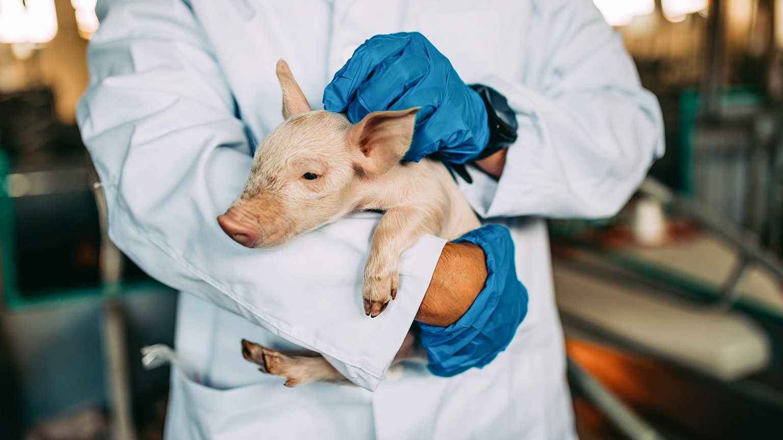
When assessing an animal’s health, there are two main types of examination:
| Distance Assessment (hands-off) | Physical Examination (hands-on) |
|---|
During a distance assessment, you observe the animal’s temperament, demeanour and behaviour as well as look for visible signs of health or health issues, including:
- assessing the animal’s breathing by listening to and watching the animal breathe, asking yourself questions such as:
- Is breathing easy or laboured?
- Are the breaths deep, shallow or irregular?
- Can you hear choking, whistling, gasping, excessive coughing, wet noises or other strange sounds while the animal breathes?
- looking for any obvious signs of bleeding, broken bones, vomiting or unusual evacuation (diarrhoea)
- looking for any issues with coat condition, for example, matted or missing fur
- looking for unusual posture or movement if the animal is already standing. If you suspect broken bones or spinal damage, do not encourage the animal to stand or move.
Demeanour is the general, observable behaviour and alertness of an animal. An animal’s demeanour is often an indication of injury, illness or disease.
Observe the animal’s behaviour and note its demeanour. The following terms are commonly used to indicate the demeanour:
- BAR - Bright, alert and responsive
- QAR - Quiet, alert and responsive
- Altered mentation
- Neurological
- Seizure
- Post-ictal – phase immediately following a seizure
- Depressed or dull
- Obtunded – lethargic or sleepy
- Stuporous – unconscious or very difficult to rouse
- Comatose.
You should conduct a distance assessment before approaching and capturing an animal and at the start of a health assessment. The particular demeanour, behaviour of the animal, and any external signs of injuries should influence your choices regarding how to approach, capture, handle, and restrain that animal.
A physical examination of an animal involves checking the animal’s vital signs and external features as indicators of general health and well-being.
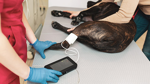
Vital signs are measurable indicators of the body’s most basic and critical functions – they are indicators of life. As part of a physical examination, five key vital signs are measured to assess the health of an animal.
- Heart rate (HR)
- Pulse rate
- Respiratory rate (RR)
- Mucous membrane (MM)
- Temperature
Collectively, temperature, pulse rate and respiratory rate are known as TPR.
Normal range of vital signs
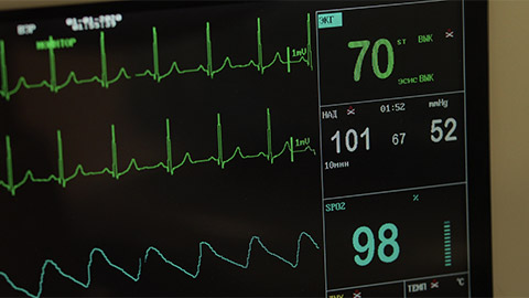
For each vital sign, there is a range of values that are considered normal for a healthy individual of that species. Vital signs that are outside the normal range tend to indicate health problems.
TPR values will vary between different species and individual animals. Variances occur between young, adult, very old, healthy or sick animals. Therefore, it is important that you can identify normal values for each species and take any relevant environmental factors into account as possible reasons for deviations from the normal.
The following table lists the normal ranges for temperature, pulse rate and respiratory rate in seven species (Cooper, Mullineaux & Turner (eds.) 2011). In Australia, temperature is measured in degrees Celsius (°C), pulse rate is measured in beats per minute (bpm), and respiratory rate is measured in breaths per minute (bpm).
| Species | Dogs | Cats | Horses | Rats | Ferrets | Rabbits | Guinea pigs |
|---|---|---|---|---|---|---|---|
| Temperature (°C) | 38.3-39.2 | 38.2-38.6 | 37.2-38.9 | 35.9-38.0 | 37.8-40 | 38.5-40 | 37.2-39.5 |
| Pulse rate (bpm) | 70-140 | 100-200 | 30-40 | 310-500 | 200-250 | 130-325 | 230-380 |
| Respiratory rate (bpm) | 10-30 | 15-30 | 12-20 | 70-115 | 33-36 | 30-60 | 90-150 |
Vital signs outside the normal range
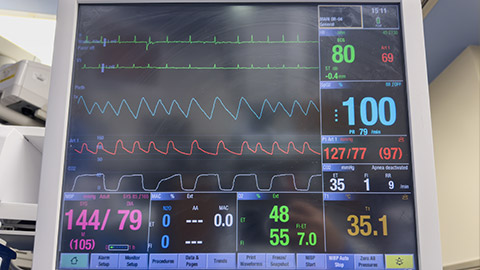
Several physiological and environmental factors affect an animal’s TPR values, such as sleep or activity. Very young animals also often have increased TPR values compared to adults of the same species.
It is advisable that if the TPR values are outside their normal ranges, the TPR should be assessed again after the environmental effects of the animal have diminished.
Frequent monitoring of vital signs can also help you assess any change in the animal’s condition – whether they are progressing (improving), deteriorating (getting worse) or remaining the same.
Heart rate and pulse rate
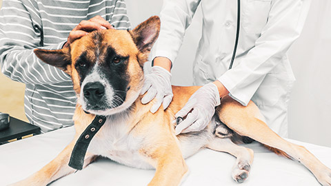
Heart rate (HR) is the number of times the heart beats in one minute. Heartbeats can be felt at several points around the body as a pulse. The number of pulses per minute is the pulse rate. In a healthy animal, the HR and pulse rate should be the same.
| Heart Rate | → | Pulse Rate |
Heart rate
To take the HR using a stethoscope:
- If required, obtain assistance to restrain the patient.
- Place the stethoscope over the left side of the chest – the best access is usually over the rib cage, just behind the front left leg.
- Listen carefully and count the number of the thumps over 15 seconds then multiply by 4 to calculate beats per minute (bpm).
- Record this information.
To take the HR without a stethoscope:
- Place your hand over the left side of the animal’s chest. Depending on the side of the animal you can use your whole hand or just your middle and index fingers. Be careful not to use your thumb. You have a strong pulse at the tip of your thumb, which can make it difficult to detect the heart rate of the animal.
- Feel for the heartbeat.
- Once you have a good hand position, count the number of the thumps over 15 seconds then multiply by 4 to calculate bpm.
- Record the HR.
The following video, ‘Merle hops over for a heart check’ (1:11 min), is an excellent demonstration of minimal, or in this case zero, restraint of an animal while conducting a vital sign check.
Pulse rate
The pulse can be measured on any part of the body where a major artery runs close to the surface of the skin. Each pulsation corresponds with a contraction of the heart. If you do not have access to a stethoscope and have difficulty finding the heartbeat with your hands, you can take a pulse rate.
The two easiest places to find a pulse in most animals are at the femoral artery (1) and the dorsal pedal (2).
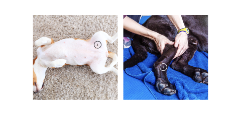
To find the femoral artery, run your hand along the inside of the hind leg until you are almost to the point the leg joins with the body. You will feel a slight dip where the femoral artery is closest to the skin. The artery feels like a thin cord between two muscles.
A pulse is generally stronger the closer it is to the heart. So, the femoral artery is usually easier to locate than the dorsal pedal pulse. However, if you can’t access the femoral artery, you can try the dorsal pedal pulse.
To find the dorsal pedal pulse, place two fingers (not your thumb) on the top (dorsal surface) of the hind foot or on the bottom (plantar) surface of the foot, just below the main pad. You need to apply a little more pressure to feel this pulse point.
Once you locate one of the pulse points, use your fingertips (not your thumb) to press down gently and feel for a pulse. As with taking the heart rate, count the number of pulses over 15 seconds then multiply by 4 to calculate bpm.
HR outside the normal range
It is important to understand what a normal HR is for the particular species and breed of animal you are working with. The size and age of the animal may also affect the ‘normal range’.
A higher than normal HR (tachycardia) could result from stress, a heart condition, anaphylaxis, blood loss or shock.
A lower than normal HR (bradycardia) may indicate many things including a heart condition, congenital disorder, poisoning or hypothermia. Some medications also lower HR.
Respiratory rate
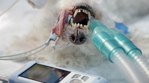
Respiratory rate (RR) is the number of breaths an animal takes in a minute. To check the patient’s RR, watch the chest rise and fall or listen closely to the animal’s breathing. Count the number of breaths for a period of 15 seconds and multiply this number by 4.
You can assess the patient’s breathing at the same time as you take the RR during your distance assessment.
RR outside the normal range
Heat stroke, airway obstruction, fluid build-up in the lungs and some heart problems are common health issues that can cause a significant increase in RR.
Medication or anaesthetic overdose, apnoea (temporary pauses in breathing, common during sleep) and some toxins and poisons will cause a significant decrease RR.
Mucous membranes
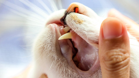
Mucous membranes (MM) are a type of tissue that line body cavities, such as the mouth. They typically contain a dense network of capillaries, and so are a good indicator of blood flow and of how much oxygen is circulating in the blood of an animal.
You can assess the MM of an animal by checking the colour of its gums.
- Moist and pink – normal and healthy
- Pale – may indicate shock or reduced blood flow
- Yellow, also known as jaundice – may indicate problems with liver function
- Blue – cyanotic (lack of oxygen) – may indicate poor breathing quality or poor circulation
- Bright red – may indicate poisoning or heatstroke.
Review the following video, ‘Checking your pet's mucous membrane colour’ (3:01 min), for a demonstration of how to check the MM of a dog. The process is essentially the same for all mammals.
Capillary refill time
Blood flows continually around the body. The speed at which it flows can be measured using capillary refill time (CRT). To measure CRT, gently compress small blood vessels (capillaries) near the surface of the skin to stop the blood from flowing through. When you release the pressure, blood will start to flow again. The time taken between lifting the pressure and blood refilling the capillary is the CRT.
One of the most common places to check CRT is on the gum of an animal, as long as it is safe to put your finger in the animal’s mouth. CRT should only take 1-2 seconds in a healthy animal. Longer than this may indicate dehydration, low blood pressure, circulatory problems or blood loss.
To check the CRT:
- Lift the animal’s lip.
- Apply a small amount of pressure with the tip of one finger to the side of the gum. As you press down, the blood leaves the area and makes the gum appear pale - blanches.
- Remove your finger and observe how long it takes for the gum to return to a pink colour.
You can assess CRT at the same time as you check the MM.
Temperature
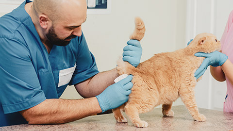
The most accurate method of taking a patient’s temperature is with a digital rectal thermometer.
Note: Taking temperature with a rectal thermometer is the most invasive technique for checking vital signs. The discomfort will increase the animal’s HR and RR. So, it should be the last vital sign you check during the health assessment.
Taking an animal’s temperature
Many animals will require some form of restraint when having their temperature taken. As always, use the minimal amount of restraint required for that animal. Ideally, one person should hold the animal’s head while a second person should take the animal’s temperature. Always lubricate the thermometer to make the procedure more comfortable for the animal.
To take an animal’s temperature using a rectal thermometer:
- Place a clean probe cover on the thermometer and lubricate the bulb.
- Lift the tail and gently insert the thermometer into the rectum, slightly rotating it as you go. Do not force entry. Gently push the thermometer against the wall of the rectum – you are taking the temperature of the animal, not its faeces!
- Keep the thermometer against the wall of the rectum for a minimum of 60 seconds (or until you hear the beep sound on some thermometers). For large animals, ensure the thermometer stays in contact with the wall of the rectum for a correct reading. Most digital thermometers will beep when you turn them on, and beep again when it’s ready to read – after maintaining a constant temperature for several seconds.
- Gently remove the thermometer. Immediately read the temperature and record the finding.
- Dispose of the probe cover appropriately, wipe away any faeces and wash and sterilise the thermometer before using it again.
Temperatures outside the normal range
When an animal’s temperature is significantly lower than the normal range, the animal is said to be hypothermic. Hypothermia can be caused by prolonged exposure to cold conditions, some diseases, blood loss and even anaesthetic.
Symptoms of hypothermia include:
- In mild cases:
- shivering
- stiff or clumsy movement
- pale gums
- confusion.
- In severe cases:
- no shivering
- rapid decrease in body temperature
- slow breathing
- fixed and dilated pupils
- collapse or lack of responsiveness (Coates 2021).
When temperatures are higher than normal, the animal is hyperthermic. A high temperature is a common indication of infection. Hyperthermia can also be caused by extreme environmental heat, such as being locked in a car on a hot day or by excessive activity on a hot day.
Hyperthermia can cause serious damage to the body and quickly lead to death if not treated.
- Early symptoms of hyperthermia include:
- Panting
- Drooling
- Bright red MM.
- Symptoms of severe hyperthermia include:
- Seizures
- Spontaneous bleeding
- Extreme lethargy
- Collapse
- Coma and death (Jones 2022).
Knowledge check 9
Refer to the table of normal ranges and identify if the animal is outside the normal range for Questions 1 to 4.
| Species | Dogs | Cats | Horses | Rats | Ferrets | Rabbits | Guinea pigs |
|---|---|---|---|---|---|---|---|
| Temperature (°C) | 38.3-39.2 | 38.2-38.6 | 37.2-38.9 | 35.9-38.0 | 37.8-40 | 38.5-40 | 37.2-39.5 |
| Pulse rate (bpm) | 70-140 | 100-200 | 30-40 | 310-500 | 200-250 | 130-325 | 230-380 |
| Respiratory rate (bpm) | 10-30 | 15-30 | 12-20 | 70-115 | 33-36 | 30-60 | 90-150 |
Case Study
At Happy Paws Veterinary Clinic, a 6-year-old domestic shorthair cat named Whiskers has been admitted for emergency surgery. Whiskers was diagnosed with a gastrointestinal blockage after showing symptoms of vomiting, lethargy, and loss of appetite. The veterinary team, consisting of Dr. Sarah, the lead veterinarian, and Lisa, the veterinary nurse, are preparing Whiskers for surgery. As part of the preparation and throughout the procedure, they will closely monitor his vital signs to ensure his safety and respond to any changes.
Initial Examination: Pre-SurgeryBackground on Whiskers:
|
|
Vital Sign Monitoring Begins: Before surgery, Dr. Sarah and Lisa conduct a thorough examination of Whiskers to establish baseline vital signs:
|
Dialogue: Pre-Surgery Preparation
 |
Dr. Sarah: "Lisa, let’s make sure we monitor Whiskers’ vital signs closely. His heart rate is slightly elevated, probably from stress, but his other vitals look good. I’ll be checking the blockage, but if anything changes, let me know immediately." |
|
| Lisa: "Got it. His respiratory rate and CRT are normal, but I’ll keep an eye on his heart rate. I’ll set up the monitors so we can track everything throughout the procedure." |
 |
|
|
During Surgery: Vital Sign Changes As the surgery begins, Whiskers is placed under anaesthesia, and Lisa continuously monitors his vital signs, using both manual checks and electronic equipment. However, partway through the procedure, Lisa notices a change in his heart rate.
|
||
| Lisa (to Dr. Sarah): "Dr. Sarah, Whiskers’ heart rate has increased to 180 bpm, and his CRT is right on the edge at 2 seconds. His mucous membranes are also starting to look a bit pale." |
 |
|
 |
Dr. Sarah: "Okay, thanks for letting me know. That increase in heart rate and delayed CRT suggests his blood pressure might be dropping. Let’s give him a fluid bolus to stabilise his circulation, and we’ll keep him warm to prevent hypothermia. We’ll need to watch his temperature too." |
|
|
Intervention: Stabilising Whiskers’ Vitals Lisa administered intravenous fluids to improve Whiskers' circulation and adjusted his position on the surgery table to ensure he’s staying warm.
|
||
| Lisa: "His heart rate is back to 160 bpm, and his mucous membranes are looking much better. CRT is improving too." |
 |
|
 |
Dr. Sarah: "Great, that’s exactly what we wanted. Let’s keep monitoring him closely as we finish the surgery. We’ll keep the fluids going and make sure his temperature doesn’t drop any further." |
|
|
Post-Surgery: Recovery and Monitoring After successfully removing the blockage, Whiskers is moved to the recovery area. The veterinary team continues to monitor his vital signs as he comes out of anaesthesia, ensuring there are no complications.
|
||
 |
Dr. Sarah: "Whiskers is waking up nicely. His vitals are stable, and we’ll continue to monitor him over the next few hours. We’ve got him on fluids to support his recovery and prevent dehydration." |
|
| Lisa: "Looks like everything’s in order. His heart rate is calming down now that the surgery is over, and his breathing is nice and steady." |
 |
Key Vital Sign Observations:
- Temperature: Whiskers' body temperature was stable, but the team took steps to prevent hypothermia, which is common during anaesthesia.
- Heart Rate: His heart rate increased during surgery, signalling potential issues with circulation, but fluids and careful monitoring helped stabilise him.
- Respiratory Rate: As expected, his respiratory rate slowed slightly due to anaesthesia but stayed within safe limits.
- Mucous Membrane Colour: The paleness observed mid-surgery was a key indicator of circulatory issues, which was resolved with fluid therapy.
- Capillary Refill Time (CRT): The slight delay in CRT helped identify a drop in blood pressure, which the veterinary team addressed quickly.
Key Learning Points:
- Close Monitoring: Regularly checking vital signs during surgery is essential for identifying potential complications before they become serious.
- Early Intervention: When Whiskers' heart rate and CRT changed, the team immediately stabilised him, preventing further complications.
- Post-Operative Care: Monitoring vital signs after surgery is just as important, ensuring the animal recovers properly from both the procedure and anaesthesia.
This case study demonstrates the importance of using the five vital signs—temperature, heart rate, respiratory rate, mucous membrane colour, and capillary refill time—to guide veterinary decisions during high-risk procedures such as surgery.
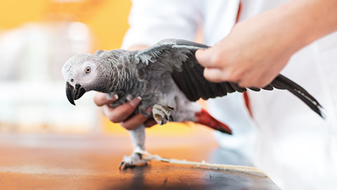
Animal general health refers to the overall well-being and physical condition of an animal. It encompasses various aspects, from the physical state of the animal’s body to its mental and emotional well-being. Regular health checks, proper nutrition, appropriate exercise, and mental stimulation are key factors that contribute to an animal's general health. Here’s a quick overview of what general health involves/ looks like:
| Physical Health | Nutrition and Hydration | Mental and Emotional Well-Being |
|---|---|---|
| Reproductive Health | Hygiene and Grooming | |
| Exercise and Fitness | ||
- Physical Health
- Appearance: A healthy animal should have a well-groomed coat or skin, bright eyes, clean ears, and a strong, clean set of teeth. Any signs of dullness, matting, discharge, or odour can indicate health issues.
- Body Condition: Maintaining an appropriate body weight is essential for general health. Obesity can lead to joint problems, diabetes, and heart issues, while being underweight could indicate malnutrition or illness.
- Movement and Activity: Healthy animals are generally active and show normal movement without signs of limping, stiffness, or lethargy. They should have an appropriate energy level for their species and age.
- Nutrition and Hydration
- Balanced Diet: Providing a diet that meets the nutritional needs of an animal is crucial for maintaining health. Nutrients like proteins, fats, carbohydrates, vitamins, and minerals should be supplied in adequate amounts, depending on the species, age, and health condition of the animal.
- Water Intake: Adequate hydration is vital for bodily functions such as digestion, circulation, and temperature regulation. Dehydration can lead to serious health problems such as kidney failure or shock.
- Mental and Emotional Well-being
- Behaviour: General health isn’t limited to physical aspects; emotional and mental well-being is equally important. A mentally healthy animal will exhibit normal behaviour for its species, whether it’s socialising with others, playing, or showing curiosity.
- Stress Indicators: Changes in behaviour, such as excessive grooming, hiding, aggression, or anxiety, can be signs of emotional distress. Enrichment activities and proper interaction can help support an animal’s mental health.
- Disease Prevention
- Vaccinations and Parasite Control: Regular vaccinations and parasite prevention (like flea, tick, and worm treatments) are essential to protect animals from infectious diseases and parasites that can impact their overall health.
- Regular Vet Check-ups: Routine vet visits allow early detection of illnesses, ensuring that any health concerns are managed before they become serious. Dental care, regular blood tests, and physical exams are part of maintaining good health.
- Reproductive Health
- Spaying/Neutering: Sterilisation helps control the animal population and can reduce the risk of certain cancers or behavioural problems. For breeding animals, ensuring proper care during pregnancy and birth is crucial.
- Hormonal Balance: Unhealthy reproductive systems can lead to conditions such as pyometra (infection of the uterus in females) or prostate issues in males.
- Hygiene and Grooming
- Cleanliness: Regular bathing, grooming, and dental hygiene are vital for maintaining the general health of animals, particularly those prone to skin conditions or dental diseases.
- Environmental Hygiene: Keeping an animal’s living space clean, with fresh bedding, clean water, and a comfortable environment, is essential for their health and well-being.
- Exercise and Fitness
- Physical Activity: Regular exercise is important for muscle tone, cardiovascular health, and mental stimulation. It can also prevent obesity and associated diseases like arthritis or diabetes.
- Age-appropriate Activities: The type and intensity of exercise should be tailored to the animal’s species, age, and health status. For example, a young dog may need more vigorous activity than an older one.
- Signs of Good General Health
- Alertness: A healthy animal is usually responsive to its surroundings, shows interest in its environment, and reacts appropriately to stimuli.
- Appetite: Regular eating habits with no significant changes in appetite are a good sign of general health.
- Normal Waste Production: The quality and frequency of an animal’s urine and faeces can reflect its general health. Diarrhoea, constipation, or changes in urination patterns can signal health issues.
- Signs of Poor General Health
- Lethargy or Weakness: Unusual tiredness or reluctance to move may indicate pain, illness, or emotional distress.
- Weight Loss or Gain: Sudden changes in weight can be a sign of metabolic issues, malnutrition, or other health concerns.
- Changes in Behaviour: Withdrawal, aggression, or altered behaviour can point to stress, illness, or pain.
- Physical Symptoms: Signs like coughing, vomiting, diarrhoea, discharge from the eyes or nose, and changes in breathing patterns often indicate health problems.
Checking the vital signs of an animal will give you a broad idea of the animal’s health. Vital signs can change quickly in response to the animal’s activity or emotional state. So, you may need to take the animal’s vital signs repeatedly to see if they are stable. If the vital signs are outside their normal ranges for a prolonged period, it may indicate something is wrong.
However, minor health and maintenance issues may not be detected while checking vital signs. Instead, a physical examination to identify external health features will help you determine the overall health status of the animal.
You do not generally need specialist equipment to conduct a physical examination of an animal. In most cases, what you can see, hear and feel is enough to identify health problems that require treatment.
Low-stress handling, restraint and examination
As with approaching, capturing and handling animals, it is best for the animal to use low-stress methods. Minimising the stress an animal experiences will also make them easier to examine.
When examining an animal try to:
- use the minimal amount of restraint possible; ideally, just use your hands
- use the minimal amount of force possible
- make the animal feel physically supported at all times
- avoid startling the animal
- avoid loud or sudden noises, including minimising environmental noise
- avoid sudden movement.
Watch
Review the following video (1:38 min) where Dingo Keeper, Lauren Nankervis, demonstrates the basic health checks she conducts on dingoes. Consider how animal training acts as a form of restraint.
Body condition score
| BCS Scales | |
|---|---|
| 1 to 5 Scale: Commonly used for farm animals (e.g., cattle, horses) |
1 to 9 Scale: Often used for companion animals (e.g., dogs, cats) |
|
|
A body condition score (BCS) is an indication of the body fat level of an animal relative to its size. BCS provides an indication of overall health in terms of nutritional status and healthy body weight.
There are two scales typically used for BCS:
- 1-5: Where a score of 1 is severely underweight; 3 is ideal weight; 5 is severely overweight
- 1-9: Where a score of 1 is severely underweight; 5 is ideal weight; 9 is severely overweight.
To determine the BSC of an animal, you need to observe the animal from above and from the side, as well as feel the animal around the ribs, backbone and hips. From above, you should see a clear waistline, and from the side, you should see the abdomen slant upward from the lowest part of the ribcage towards the hind legs. When you feel the animal along its backbone and across the ribs and pelvic bones, you should be able to feel the bones through the body fat with only light pressure. However, the bones should not be visible through the skin (Williams & Buzhardt n.d.).
Assigning a BCS is subjective. However, there are pictorial charts for many animal species to help guide the assessor, such as those provided by The World Small Animal Veterinary Association (WSAVA):
- Body Condition Score (pdf) (chart for cats)
- Body Condition Score (pdf) (chart for dogs).
How is BCS Assessed
BCS is determined through both visual inspection and palpation (feeling the animal’s body, particularly around the ribs, spine, hips, and tail). The examiner checks for fat deposits and muscle mass to evaluate the animal's condition.
- Underweight animals: Ribs, spine, and hip bones are often prominently visible, and there’s little to no fat coverage over these areas. Muscle mass may also be depleted.
- Ideal weight animals: The ribs can be felt but not easily seen, and there is a thin layer of fat over the body. The animal has a healthy muscle tone and a defined waist.
- Overweight/obese animals: Ribs and spine are difficult to feel under a thick fat layer. Fat deposits are noticeable on the back, tail base, and abdomen. The waist is not visible, and the body may appear rounded or distended.
Importance of BCS
- Nutritional Management: BCS is critical for determining if an animal is receiving the right amount of food. Adjusting feed based on BCS helps in maintaining a healthy weight, improving performance, and preventing diet-related diseases.
- Health Monitoring: A consistently high or low BCS can indicate underlying health problems. Underweight animals may suffer from malnutrition, parasites, or illness, while overweight animals are at risk of conditions such as diabetes, joint problems, or heart disease.
- Breeding Animals: In breeding stock, maintaining an appropriate BCS is important for reproductive performance. Underweight animals may have fertility issues, while overweight animals may have difficulty giving birth or suffer from metabolic problems.
BCS Example of a Dog (1-9 scale)
| 1 | BCS 1: Emaciated—Ribs, lumbar vertebrae, and pelvic bones are visible from a distance; no palpable fat. |
| 2 | BCS 3: Thin—Ribs are easily visible; minimal fat cover. |
| 3 | BCS 5: Ideal—Ribs can be felt without excess fat; the waist is visible when viewed from above, and the abdomen is tucked when viewed from the side. |
| 4 | BCS 7: Overweight—Ribs are difficult to feel under fat; slight abdominal rounding. |
| 5 | BCS 9: Obese—Ribs cannot be felt under a thick fat layer; heavy fat deposits over the lumbar area and base of the tail; the abdomen is distended. |
Examples of BCS Score in different animals
Here are ten Body Condition Score (BCS) examples across different pets, from very thin (BCS 1) to obese (BCS 9). Each example will include observable signs explaining why the pet has that particular score.
1. Dog - BCS 1 (Emaciated) |
|
|---|---|
2. Cat - BCS 2 (Very Thin) |
|
3. Rabbit - BCS 3 (Thin) |
|
4. Horse - BCS 1 (Underweight) |
|
5. Dog - BCS 5 (Ideal) |
|
6. Cat - BCS 6 (Slightly Overweight) |
|
7. Guinea Pig - BCS 7 (Overweight) |
|
8. Horse - BCS 8 (Obese) |
|
9. Dog - BCS 9 (Morbidly Obese) |
|
10. Cat - BCS 9 (Morbidly Obese) |
|
Animal examination techniques
Animal examination techniques involve a series of steps and methods used by veterinarians and animal care professionals to assess the overall health of an animal. These techniques provide insight into the animal's physical condition, detect signs of illness or injury, and guide diagnosis and treatment. A comprehensive examination includes observing, palpating, and listening to various parts of the animal's body, as well as taking measurements such as vital signs.
To give an animal an overall physical examination, it is best to work systematically so you don’t accidentally skip something. The key external features to examine are:
| Hydration | Eyes and Nose | Mouth | Ears | Lymph Nodes |
| Lungs | Abdomen | Anus | Limbs | Skin |
Follow the advice of your supervisor regarding the order in which to carry out the health checks. Monitor the animal’s behaviour throughout the examination. If any of the techniques cause the animal discomfort or pain, you may be at risk of being bitten. Adjust your level of restraint throughout the examination in response to any changes in the animal’s behaviour or temperament.
Hydration level
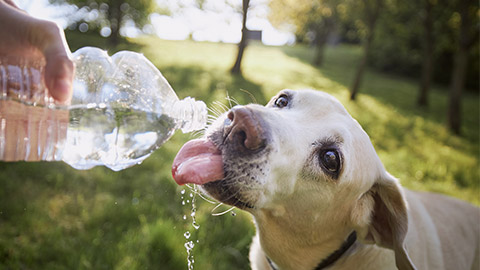
The hydration status of an animal is an indication of the amount of water present in the body. Water is essential for most bodily processes as well as for maintaining good blood pressure. Dehydration occurs when the animal can’t (or won't) drink enough or if they lose water through repeated vomiting or diarrhoea.
Dehydration can cause organ damage and even death in severe cases. Dehydration is particularly dangerous in small and young animals, such as puppies and kittens, because it can happen very quickly.
There are two common ways to assess the hydration status of an animal:
| The skin tent test | The gum test |
|---|
Skin tent test
To conduct a skin tent test (also known as the skin turgor test):
- Gently pinch and lift the skin between the shoulder blades, forming a ‘tent’ shape.
- Drop the skin.
In a well-hydrated animal, the skin should spring straight back to its normal position after you release it. If the skin returns slowly or remains in the tented position, it indicates dehydration. The longer the skin takes to return, the more severe the dehydration.
Gum test
To conduct a gum test:
- Gently lift the animal's upper lip and look at the gums.
- Use your finger to touch the surface gently.
In well-hydrated animals, the gums should look glossy and wet. In dehydrated animals, the gums tend to look dull and are sticky to the touch (Bond Vet 2020).
Eyes and nose
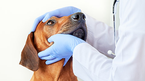
Look closely at the animal’s eyes and nose.
Eyes
The eyes should appear moist, clear and bright. The whites of the eyes should appear white with only a few visible blood vessels. The pupils should be the same size, should contract equally when exposed to a bright light and expand when the room is darkened (Gfeller, Thomas & Mayo 2020).
Abnormal signs to check for include:
- unusual or excessive discharge
- tear staining (fur around the eyes is stained pink)
- inflammation (redness or swelling) of eyelids
- pupils that do not respond normally to changes in light conditions
- colouration of the whites of the eyes.
Nose
A common misconception is that healthy animals should have a ‘wet nose’. This is not true.
Instead, check the nose for unusual or excess discharge, cracks and bleeding.
Mouth
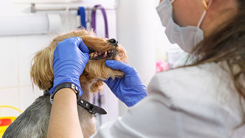
As always, only check an animal’s mouth and teeth if it is safe to do so.
A healthy mouth should have moist pink gums and clean, white teeth. When checking an animal’s mouth note:
- bad breath
- plaque or tartar on the teeth (usually around the base of the teeth)
- inflammation of the gums (periodontal disease)
- missing or broken teeth.
Ears
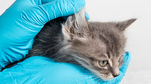
Ear shape and posture vary greatly between species and breeds. However, all healthy ears should be clean and dry, have little-to-no odour and be pain-free when touched.
Signs of health issues in the ears include:
- inflammation
- discharge
- strong smell
- erythema (skin irritation and redness)
- pain or tenderness
- wounds, scabs, lumps or rashes on the skin
- signs of external parasites.
Lymph nodes
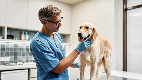
Lymph nodes are a key part of the immune system. They are sites around the body where white blood cells gather and fight pathogens. Swollen or painful lymph nodes are a common sign of infection.
The largest and easiest lymph nodes to locate on a mammal, especially for cats and dogs, are found in the neck, chest, armpits, groin and behind knees (Dogtor Pete 2022).
Gently palpate (feel around carefully with your fingers) the areas where the lymph nodes are located. If they are swollen, you should feel hard lumps quite easily. However, if you’re unsure what it is you are feeling for, it is easy to mistake other lumps (such as swollen salivary glands) for swollen lymph nodes. If you find a lump that you suspect is a swollen lymph node, ask your supervisor or a vet to check.
Lungs
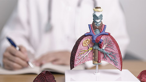
You will have assessed the animal’s breathing during your distance assessment. However, if you have access to a stethoscope, you can use it to listen to the lungs and detect quieter sounds of breathing difficulties.
Abdomen
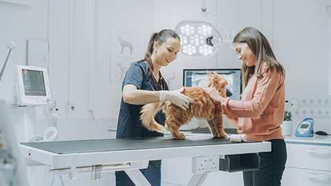
Gently palpate the abdomen (stomach area), starting just behind the ribs and working your way down to the pelvic bone. Feel for lumps, bumps and masses, tightness and bloating. If the animal has difficulty breathing or shows signs of discomfort or pain during the process, it could indicate a serious problem (Gfeller, Thomas & Mayo 2020).
Anus
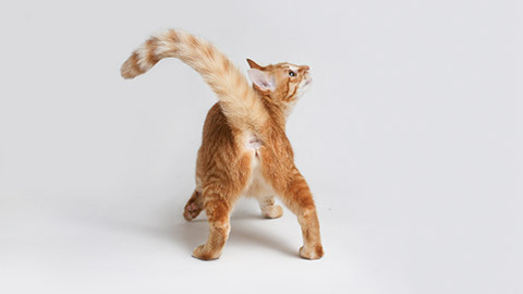
Life the tail, if necessary, to examine the animal’s anus. Check for faecal staining, which could indicate diarrhoea, and for enlarged anal glands, which may indicate a blockage.
Limbs
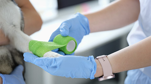
Examine each limb, starting at the foot. Check:
- claws/nails/hooves for:
- damage or absence
- overgrowth
- discolouration.
- foot pads for:
- lodged grass seeds or other foreign bodies
- injuries
- swelling or inflammation.
- for broken bones
- for range of motion through each joint.
Skin

Finally, skin condition can be a good indicator of the overall health of the animal. Run your hands over the whole of the animal to check for abnormalities, including:
- wounds
- missing fur/hair or poor coat condition
- lumps, bumps or masses
- excessive oil or discharge
- unpleasant odour
- foreign objects and ectoparasites, such as ticks.
The following video, ‘How To Perform A Clinical Exam On Your Dog | Vet Demo’ (10:57 min), provides a full demonstration of a basic health check on a dog. You are not expected to conduct a full examination by yourself. However, it is in your best interest to observe the demonstration, so you are more familiar with the process and can better assist the vet or other staff member conducting the examination.
Knowledge check 10
Case Study
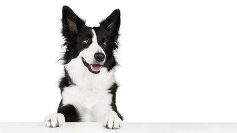
A 5-year-old Border Collie named Max is brought to the vet clinic for a routine general health check. Max’s owner, Mrs. Anderson, reports that Max has been eating well, is active, and hasn’t shown any significant behavioural changes. The vet, Dr. Lewis, begins a full physical examination to assess Max’s overall health and check for any underlying issues.
Step 1: Observation (Visual Inspection)Dr. Lewis starts by observing Max from a distance before making contact, allowing her to assess the dog’s overall appearance, posture, behaviour, and movement. Max seems bright, alert, and responsive to his surroundings, showing no signs of anxiety or distress.
Outcome: Max’s coat is healthy and shiny, and his movement is normal with no signs of lameness. His behaviour is alert and curious, which is typical for his breed. |
Step 2: Palpation (Feeling the Body)Dr. Lewis moves on to palpation, where she gently feels different parts of Max’s body to check for any abnormalities beneath the surface.
Outcome: Max’s lymph nodes are normal in size, and his abdominal organs feel smooth with no abnormal masses or signs of pain. His muscles and joints feel strong, and there is no discomfort during palpation. |
Step 3: Auscultation (Listening to the Body)Next, Dr. Lewis uses her stethoscope to listen to Max’s heart and lungs, focusing on any irregularities in sound or rhythm.
Outcome: Max’s heart rate is steady, and there are no murmurs or irregular sounds. His lung sounds are clear, with no indication of respiratory problems. Gut sounds are present and normal, indicating proper digestion. |
Step 4: Temperature MeasurementDr. Lewis then measures Max’s body temperature using a rectal thermometer to check for signs of fever or hypothermia.
Outcome: Max’s temperature is 38.7°C (101.7°F), which is within the normal range. |
Step 5: Mucous Membrane and Capillary Refill Time (CRT) CheckDr Lewis examines Max’s gums (mucous membranes) and performs a capillary refill test by pressing gently on the gums and checking how quickly the colour returns.
Outcome: Max’s gums are pink, and the capillary refill time is normal, indicating good circulation and oxygenation. |
Step 6: Weight and Body Condition Score (BCS)Dr. Lewis weighs Max and evaluates his Body Condition Score (BCS) to ensure he is maintaining a healthy weight. She also feels for the amount of fat over Max’s ribs and hips.
Outcome: Max weighs 22 kg, which is consistent with his last check-up. His BCS is 5, indicating he is in ideal condition—ribs can be felt without excess fat, and Max has a visible waist when viewed from above. |
Step 7: Hydration Status (Skin Turgor)Dr. Lewis checks Max’s hydration by performing a skin turgor test. She gently pinches the skin on Max’s neck to see how quickly it returns to normal.
Outcome: Max’s skin snaps back into place immediately, indicating he is well-hydrated. |
Step 8: Oral and Dental ExaminationDr. Lewis examines Max’s teeth, gums, and oral cavity for any signs of dental disease, such as tartar build-up, gum inflammation, or broken teeth.
Outcome: Max’s teeth are in good condition, with no significant tartar build-up or gum disease. His oral cavity is healthy. |
After completing the general health exam, Dr. Lewis concludes that Max is healthy. His physical condition is ideal for his age and breed, and there are no signs of illness or abnormalities. She advises Mrs Anderson to continue with Max’s current diet and exercise routine and recommends a follow-up visit in six months for another routine check-up.
Dr. Lewis also reminds Mrs. Anderson to keep Max up to date on his vaccinations and parasite prevention treatments.
Recording health observations involves systematically documenting an animal’s physical and behavioural status during an examination or throughout care. This practice is crucial in veterinary and animal care settings to track changes in health over time, ensure continuity of care, and provide a detailed history for treatment planning. Proper recording allows for quick identification of health trends or issues and ensures that accurate information is available for all staff involved in the animal's care.
When assessing an animal’s health, it is important to maintain accurate records. This information contributes to the patient’s history and may help with diagnosis or treatments in the future.
When recording observations, it is important to include all the relevant detail while being concise.
What observations to record
The specific information you need to record will depend on your workplace and the purpose of the health check. For example, a veterinary clinic will keep different health care records from a boarding facility. If you were monitoring the ongoing health of an animal in a shelter, you might only record your observations from the distance assessment, while you may need to record repeated vital sign checks over a period of time in a first aid situation.
Follow the specific procedures for your workplace and remember to use appropriate industry terminology, abbreviations and units as appropriate. Confirm the requirements with your supervisor if you are not confident.
Some common health observations that you may need to record include:
- the weight of the animal (kg)
- body condition score (BSC)
- the amount of food/water consumed
- evidence of urination or defecation
- demeanour and behavioural observations
- distance assessment observations
- observations of coughing/sneezing
- vital signs
- other physical examination assessments.
Key Components of Recording Health Observations
|
Identification of the Animal
|
|
Date and Time
|
|
Vital Signs
|
|
Body Condition Score (BCS)
|
|
Behavioural Observations
|
|
Physical Appearance
|
|
Palpation Findings
|
|
Auscultation Results
|
|
Hydration Status
|
|
Weight and Growth Monitoring
|
|
Pain and Discomfort
|
|
Feeding and Elimination
|
|
Medications and Treatments
|
|
How to record observations
The specific method of recording observations will vary between workplaces. Some workplaces may use an app on a mobile device, others may record observations manually on a form or on a computer database. Again, follow the procedures of your workplace.
Review the article, ‘Instructions for daily monitoring of animal health and behavior’, for more information about the importance of record keeping in an animal shelter. You can view six sample record forms by following the links at the bottom of the article. Take note of the abbreviations and codes used to quickly and accurately record observations.
Industry terminology
When recording health observations, it is important to use appropriate industry-accepted terminology and abbreviations. That way when someone else reads your records, they will understand what you meant. For example, hyperthermia is the correct term compared to simply saying ‘overheating’ or ‘too hot’.
Abbreviations are an excellent way of taking notes quickly. However, don’t make up your own! Use the industry standard abbreviations. If you make up your own abbreviations or shorthand, other people will have difficulty understanding your records.
Refer to the table of abbreviations in Topic 2 of this module to refresh your memory of some common industry abbreviations.
Ongoing monitoring
In animal care facilities where animals are kept for long periods of time, such as animal shelters, boarding facilities, wildlife sanctuaries or zoos, you will likely need to monitor the ongoing health of animals. In many workplaces, basic health monitoring occurs daily with more in-depth check-ups less frequently.
Daily monitoring allows you to observe changes in the animal’s condition. The three general updates regarding animal condition are:
- Deterioration (getting worse)
- Progress (getting better)
- No change.
You should report any signs of deterioration immediately to your supervisor.
Terminology
| Unit or term | Explanation or definition |
|---|---|
| mg/L | Milligrams per litre |
| kg | Kilogram |
| g | Grams |
| Environmental Hazard | A source of potential harm to the environment |
| Personal Precaution | A measure taken to reduce the risk to yourself |
| Ocular | Eyes |
| Aquatic | Water-based/ Freshwater |
| Ectoparasite | Parasites that live on the surface of the host |
Knowledge Check 11
Explore
In this section are some examples of health observation documents with which you can familiarise yourself. Remember that all animal care services would have their own templates and processes to record information. This might be digital or in paper format.
Case Study
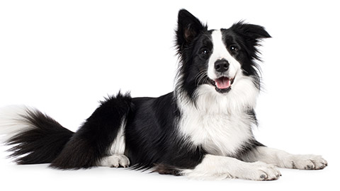
Observation Record of Max
Here is an example of an Observation about Max the Border Collie.
|
Observation Record for a Dog (Max):
|
