In this topic, we focus on the structure and function of the cardiovascular system.
The cardiovascular (CV) system is essential to every aspect of human life and athletic performance. The cardiovascular system (cardio~= heart, vascular~= blood or blood vessels) is composed of 3 components:
- The heart
- Blood vessels
- Blood
The role of the CV system is to provide essential nutrients to the body's cells and tissues when required and remove waste products from the body. We will look at the heart, blood vessels, and blood, and how they each contribute to athletic performance as well as the maintenance of homeostasis.
In this topic, you will learn about:
- The heart
- Blood and Blood vessels
- Circulatory system
- Acute measurements of heart function
- Long-term cardiovascular adaptations.
Watch
This short video shows the internal structure of the heart. Watch it to get familiar with the heart's structure before progressing.
The heart is the 'battery' of the cardiovascular system. Without the heart beating and pumping blood through our vessels, the CV systems would be unable to remove waste and deliver nutrients throughout the body.
Heart function
The heart has one primary function. For the body to stay alive, each cell must receive a continuous supply of nutrients and oxygen. At the same time, carbon dioxide and other materials produced by the cells must be transported away from the tissues and removed from the body.

Move blood heart and lungs

Move blood heart and body tissues

Pump blood around the body

Maintain homeostasis
These processes are continually maintained by the body’s cardiovascular system (circulatory system). It is the heart's job to produce the pressure required to move blood between the heart and lungs, and the heart and body tissues, pump blood around the body and maintain homeostasis.
Did you know?: Your heart beats approximately 10,000 times a day. This amounts to approximately 35 million beats in a year and 2.5 billion times in an average lifetime.
Heart structure
Your heart is roughly the same size (but not the same shape) as your fist and weighs approximately 250g for females and 300g for males.
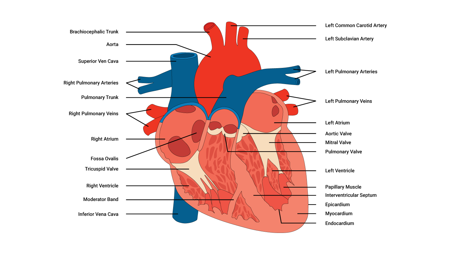
Chambers
Our heart is composed of 4 separate chambers that contribute to how it performs its pumping action. There are 2 superior receiving chambers (the atria) and 2 inferior (below the atria) pumping chambers (the ventricles), with one of each on the right and left portions of the heart.

| Right and left atria: Receiving chambers |
The right and left atria receive blood returning to the heart. The right atrium receives deoxygenated blood returning from the body tissues, while the left atrium receives oxygenated blood from the lungs. Between the right and left atria is a layer of tissue called the atrial septum that separates the left and the right receiving chambers. |
|---|---|
| Right and left ventricles: Pumping chambers |
The right and left ventricles are the heart pumps. They receive the blood from the corresponding atria via 'gateways' called valves. These valves open when there is sufficient pressure pushing against them, controlling the rate and flow of blood from the atria to the ventricles. The ventricles pump the blood to relevant parts of the body depending on the oxygen status of the blood. The right ventricle pumps deoxygenated blood to the lungs, while the left ventricle pumps oxygenated blood to the body tissues. |
Valves
4 valves separate the different chambers of the heart. They are controlled by the nervous system and work together to keep blood moving through the heart and around the body. Most importantly, they keep blood moving in the same direction.
The valves perform their function during the phases of a heartbeat.
- Systole - a contraction of the ventricles
- Diastole - relaxation of the ventricles
Coronary arteries
The heart is a specialised muscle, and like any muscle, the heart requires oxygen to stay alive. Even though our heart contains oxygen-rich blood, it cannot gain oxygen from the blood inside it. Instead, coronary arteries deliver O2 rich blood. These arteries branch off into smaller arteries that envelope the heart.
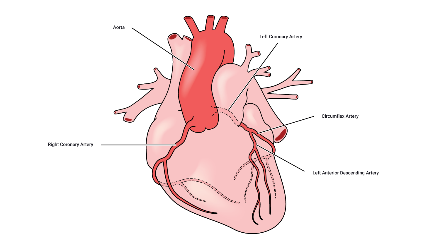
Typically, there is one artery on the right side and one on the left, with the left one generally being the larger. This is because the left side of the heart pumps blood to the body, whereas the right-hand side pumps blood to the lungs.
The left main coronary artery divides into 2 sizable branches, the left anterior descending (supplying the front of the heart) and the left circumflex (wrapping around the left side and back of the heart). The right coronary artery supplies the back of the heart.
Blockages of these vessels are one of the leading causes of death.
Muscular tissue of the heart: Myocardium
The heart is composed primarily of muscular tissue, specifically the myocardium.
When you look at a cross-section of the heart, you can see a subtle difference in the myocardium. The left-hand side of the heart (on the right), particularly around the ventricle, is substantially thicker than around the right ventricle. This is because the left ventricle pumps blood around the entire body, so it needs to be strong enough to create sufficient pressure to do so.
Heart muscle anatomy
How the heart beats
The cardiac muscles of the heart are referred to as autorhythmic fibres because they are self-excitable. This refers to the fact that they repeatedly generate their own action potentials that trigger contractions of the heart muscle fibres.
Heart conduction system
The heart conduction system is the network of nodes, cells, and signals that controls your heartbeat. Conduction follows a complex sequence that coordinates the pumping of each heart chamber at the right time. This allows blood to flow in the correct direction and with the appropriate amount of force, for example, the left ventricle must pump harder than the right to deliver blood to the body. This is controlled by your autonomic nervous system and is regulated by a system of chemoreceptors and electrical impulses.
Blood
Aside from both starting with the letter "b", how are bones and blood similar? Surprisingly, they are considered the same type of "connective tissue". Connective tissue is one of four types of tissue that make up the human body (the others include epithelium, muscle, and nervous) and has many functions. With that in mind, let us now discuss the components of blood.
Structure
Blood comprises 4 basic components: plasma (the liquid component) and 3 types of blood cells (the solid component).

| Plasma | A watery straw-coloured liquid containing ions, proteins, and hormones. It carries the blood cells around the body. |
|---|---|
| Red blood cells (erythrocytes) | These contain hemoglobin to transport oxygen. |
| White blood cells (leukocytes) | Fights infection from illness and disease. Your body makes more white blood cells to fight bacteria and viruses when you are sick. This is why you might get a white cell count when unwell. |
| Platelets (thromocytes) | Controls bleeding by clotting at wounds. |
Blood vessels
Blood vessels are the pathways for blood around the body. There are 5 types of blood vessels:
- Arteries
- Arterioles
- Capillaries
- Venules
- Veins
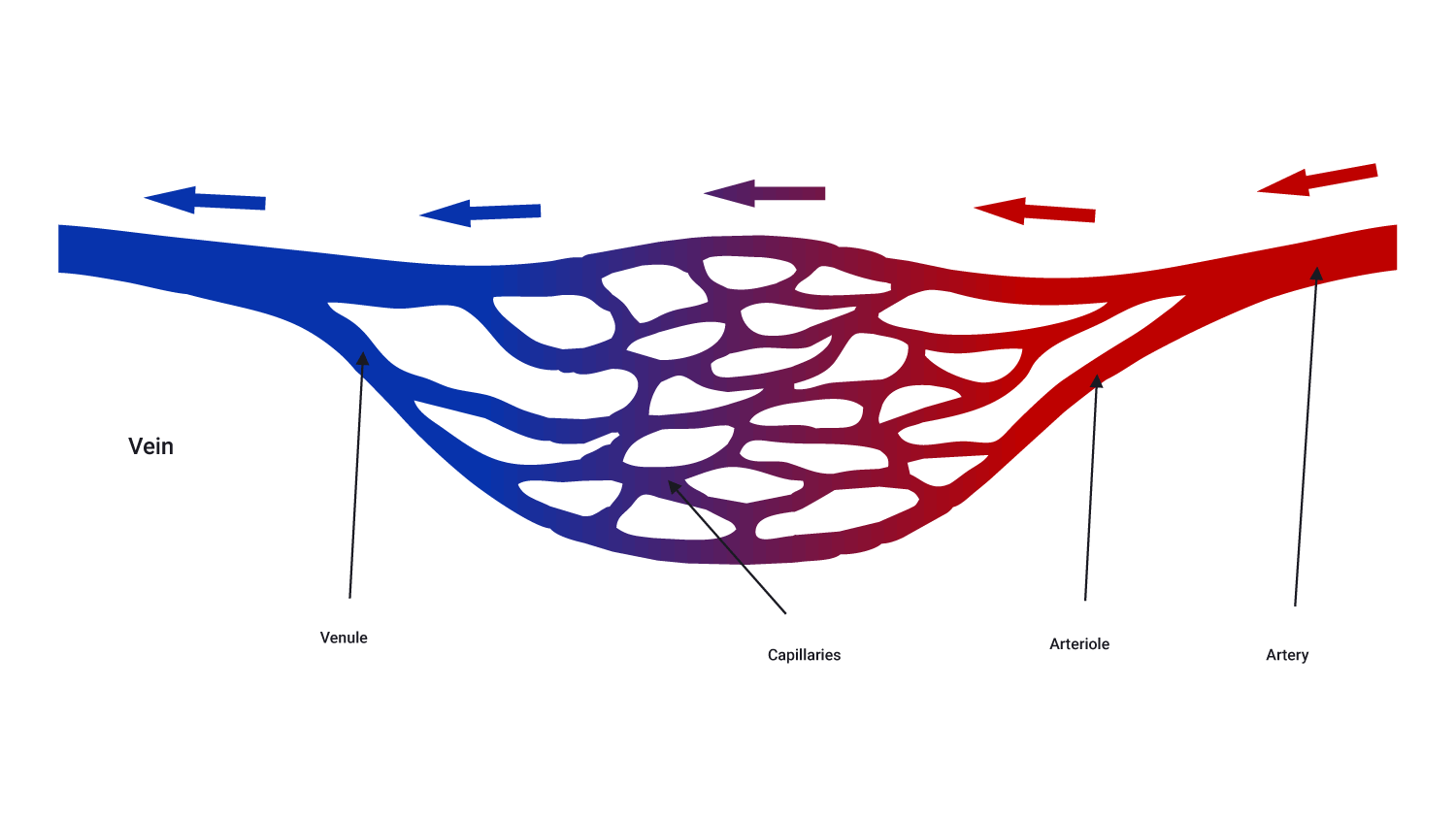
Arteries
Arteries are the largest blood vessels and carry blood away from the heart. There are 2 types:
- Systemic: Carry blood from the aorta to be distributed to the body tissues. These carry only oxygenated blood.
- Pulmonary: Carry blood from the pulmonary trunk to be distributed to the alveoli in the lungs. These carry only deoxygenated blood.
Arteries branch at intervals along their length into arterioles which are tiny branches of arteries that lead to capillaries. Along with smaller arteries, these are under the control of the sympathetic nervous system and constrict (narrow) and dilate (widen) to regulate blood flow.
Arterioles
These are small structures that deliver blood to capillaries. Arterioles are the main effectors of blood pressure.

Capillaries
Capillaries are tiny, extremely narrow, blood vessels, approximately 5-20 micro-metres in diameter. They are so tiny, that red blood cells pass through in a single file line.
There are networks of capillaries in most of the organs and tissues of the body. Capillaries are supplied with blood by arterioles and are drained by venules. Capillary walls are very thin. This allows them to exchange substances and gasses between the blood and the surrounding tissue e.g., oxygen, carbon dioxide, water, and salts.
Venules
Venules are small vessels that drain blood from capillaries and into veins. Many venules unite to form a vein. They drain blood from capillaries and pass it on into veins for return to the heart.
Veins
Veins transport blood towards the heart. There are 2 types:
- Systemic: Carry deoxygenated blood back to the right atrium of the heart.
- Pulmonary: Carry oxygenated blood from the lungs back to the left atrium of the heart.
Veins have valves that prevent blood from flowing in the reverse direction.
Blood follows a certain pattern through vessels in and out of the heart.
Deoxygenated and oxygenated blood
In anatomical diagrams, you will usually see blood depicted in two colours: blue and red.
- Blue represents deoxygenated blood. This blood lacks oxygen O2. It is heading back to the lungs to receive oxygen inhaled from the environment.
- Red indicates oxygen-rich (oxygenated) blood. This blood has been oxygenated in the lungs and is now on its way to the tissues and cells of the body.
All the body's cells need oxygen to efficiently create energy and perform tasks such as digestion, muscle contraction, and cell communication.
Take a look at the following image, which highlights the direction of travel of blood both into and out of the heart.
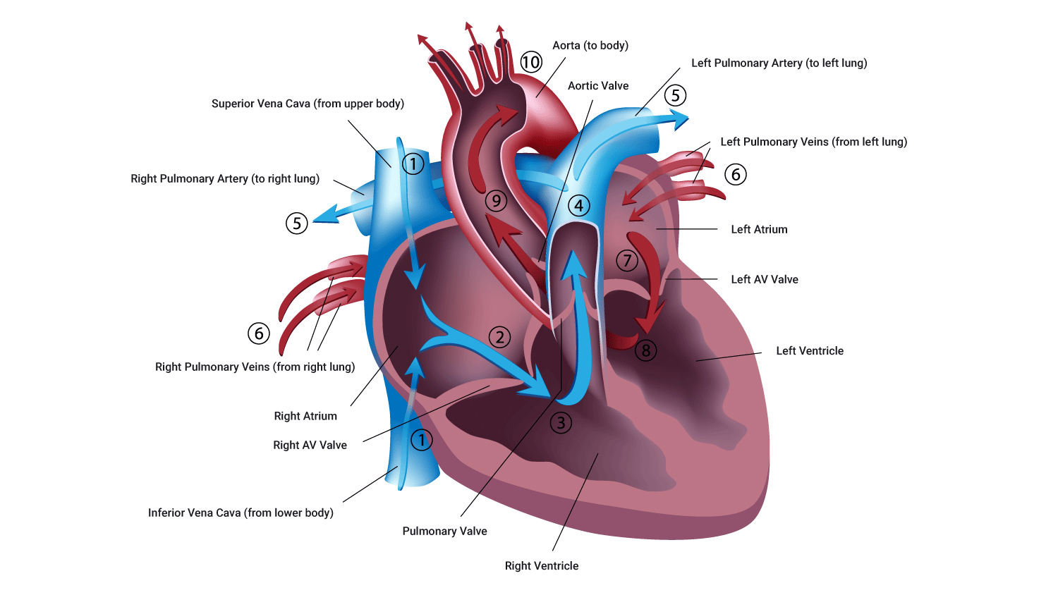
Remember that when you look at a picture of a heart front-on, the left is on the right, and the right is on the left.
The pathway of blood through the heart is of great physiological importance when working in industries such as health and fitness. Becoming comfortable with the direction of blood, and the factors involved in pumping blood through the heart and the body, will help you design the best programmes for your clients. Understanding the circulatory system and having the ability to calculate capacities suitable for your clients, are incredibly valuable tools to draw on when you design a tailored routine for your clients.
Watch
This video goes through the steps of how blood flows through the heart. It's only a couple of minutes long, but you may want to watch it a few times to get really comfortable with it.
Let's look at the pathway of blood through the heart.
Deoxygenated blood flow
This is known as the pulmonary circuit and happens on the right side of the heart.
- Deoxygenated (oxygen-poor) blood arrives in the right atrium (receiving chamber) from the superior vena cava (upper body) and the inferior vena cava (lower body).
- Blood gathers in the right atrium, which applies pressure to the atrium walls and the tricuspid valve.
- When a sufficient volume of blood arrives in the right atrium, the tricuspid valve opens, and blood then enters the right ventricle.
- When the ventricle is full, pressure is applied to the rear of the tricuspid valve, which closes the tricuspid valve. This prevents the backflow of blood while the ventricle contracts.
- The right ventricle contracts, pushing blood out of the ventricle, and up the pulmonary artery. This is enabled by the opening of the pulmonary valve.
- This deoxygenated blood then arrives, via the pulmonic artery to the lungs. Gas exchange takes place and the blood is refreshed with oxygen, and carbon dioxide is removed through exhalation.
At the same time as deoxygenated blood is flowing through the heart, oxygenated blood is also flowing through.
Oxygenated blood flow
This is known as the systemic circuit and happens on the left side of the heart.
- Oxygen-rich blood arrives via the pulmonary vein and empties into the left atrium.
- When sufficient blood is gathered here, producing sufficient pressure on the mitral valve, the mitral valve opens.
- Blood flows from the left atrium into the left ventricle via the mitral valve.
- When the left ventricle is full of blood, the pressure of this blood pushes back on the mitral valve. This forces it to close and protects against backward blood flow into the atria while the ventricle contracts.
- As the ventricle contracts, blood is propelled into the aorta via the aortic valve. It then travels into the veins and onwards to the body's cells.
Watch
Now is a great time to watch this video, it nicely sums up what we have discussed. Exhale, take a break, and enjoy the next few minutes.
Check that you have a good understanding of the cardiovascular system before moving to the next topic. You'll be assessed on the cardiovascular system in a combined test with the respiratory system.