In this topic, we explore the structure and function of the nervous system. The nervous system is responsible for tasks that help maintain homeostasis, so there is a lot of detail. Use the activities to check your understanding frequently.
The nervous system does not maintain homeostasis alone. It works with other systems to maintain a safe functioning working environment for the human body.
Think of the nervous system as the control centre. It is the receiver of all messages from the other 10 body systems and is the brains of the operation. The nervous system:
- receives messages from the other body systems.
- translates them as to what, why, where and when attention is needed and how it will perform this action in the body.
- communicates this to other systems and lets them know what they need to do.
This is teamwork!
Understanding the nervous system and its components will help you understand how muscles work in terms of exercise and when working out and cooling down. This will equip you to be confident and knowledgeable while providing your clients a safe and valuable experience.
In this topic, you will learn:
- Nerve anatomy and physiology
- The nervous system and exercise
- Nervous system adaptations.
Structure and Function
The nervous system carries out numerous functions within the human body. It enables us to speak, smell, remember past information, make new memories, and learn. It provides signals which control body posture and movements, reflexes, and regulates our internal environment.

The nervous system is divided into 3 basic functions:
- Sensory input
- Integration (processing)
- Motor output
1. Sensory input
The sensory receptors detect stimuli. They detect information such as increased body temperature or external stimuli such as a cold breeze. This sensory information is carried to the brain and spinal cord for processing through the spinal and cranial nerves.
2. Integration
This is when the nervous system analyses the sensory input and makes a decision as to how and when to respond.
3. Motor output
The nervous system sends messages (also known as motor responses) that activate organs to move and/or the glands to secrete their substances.
What does the nervous system do?
The nervous system and the endocrine system work to control all activities within the human body. The nervous system quickly communicates messages between the brain and the body using nerve impulses (also known as action potentials).
The 4 main functions of the nervous system are:
- Control of the internal environment to achieve homeostasis
- Reflexes
- Memory and learning
- Voluntary control of movement
These functions are relevant for personal trainers to understand and be aware of when programming. Let's look at these functions in a bit more detail.
Control of the internal environment to achieve homeostasis
The nervous system carries out homeostasis to ensure internal conditions stay constant. This includes maintaining blood glucose levels, body temperature, and water levels.
Receptor cells detect changes in the environment (stimuli). The central nervous system (CNS) receives and processes the information and communicates to effector cells (e.g., muscles and glands) what to do to restore the body to optimal levels.
For example, when we exercise, we create heat. To maintain a relatively constant temperature, the nervous system sends messages to blood vessels to dilate, increasing blood flow to the skin and increasing sweating. This helps disperse the heat.
Reflexes
Reflex activity is when a stimulus leads to immediate action. The action is an inborn central nervous system activity, not involving consciousness.
A well-known example of this is the pupillary light reflex. If a light is flashed near one eye, the pupils of both eyes contract. Light is the stimulus; impulses reach the brain via the optic nerve, and the response is conveyed to the pupillary musculature by autonomic nerves that supply the eye.
Reflex functions protect us from injury. If you are jogging and accidentally roll your ankle, the stretch reflex instantly protects the ankle from breaking, limiting the injury to a sprain. The instant stretch in the muscles around the ankle is the stimulus. The CNS interprets this and sends a message to the muscles to tell them to contract and resist the stretch.
Memory and learning
As you learn, messages travel from one neuron to another over and over, creating connections, or pathways, in the brain. The first time you do something, it requires a lot of concentration and effort. But with practice, the connections (or pathways) are strengthened and the task or memory recall becomes easier. The pathway becomes established.
Repetition assists learning and memorisation and this can be applied to exercise. New movements, especially complex ones, take time for the nervous system to learn.
Voluntary control of movement
Every voluntary movement a person performs is under the direct control of the nervous system. The nervous system sends messages to the targetted body parts to move. If the movement has been repeated many times, the movement will be very efficient. E.g., walking. If the movement is new, it will be less efficient and may look awkward and ungainly. E.g., a person learning to squat for the first time.
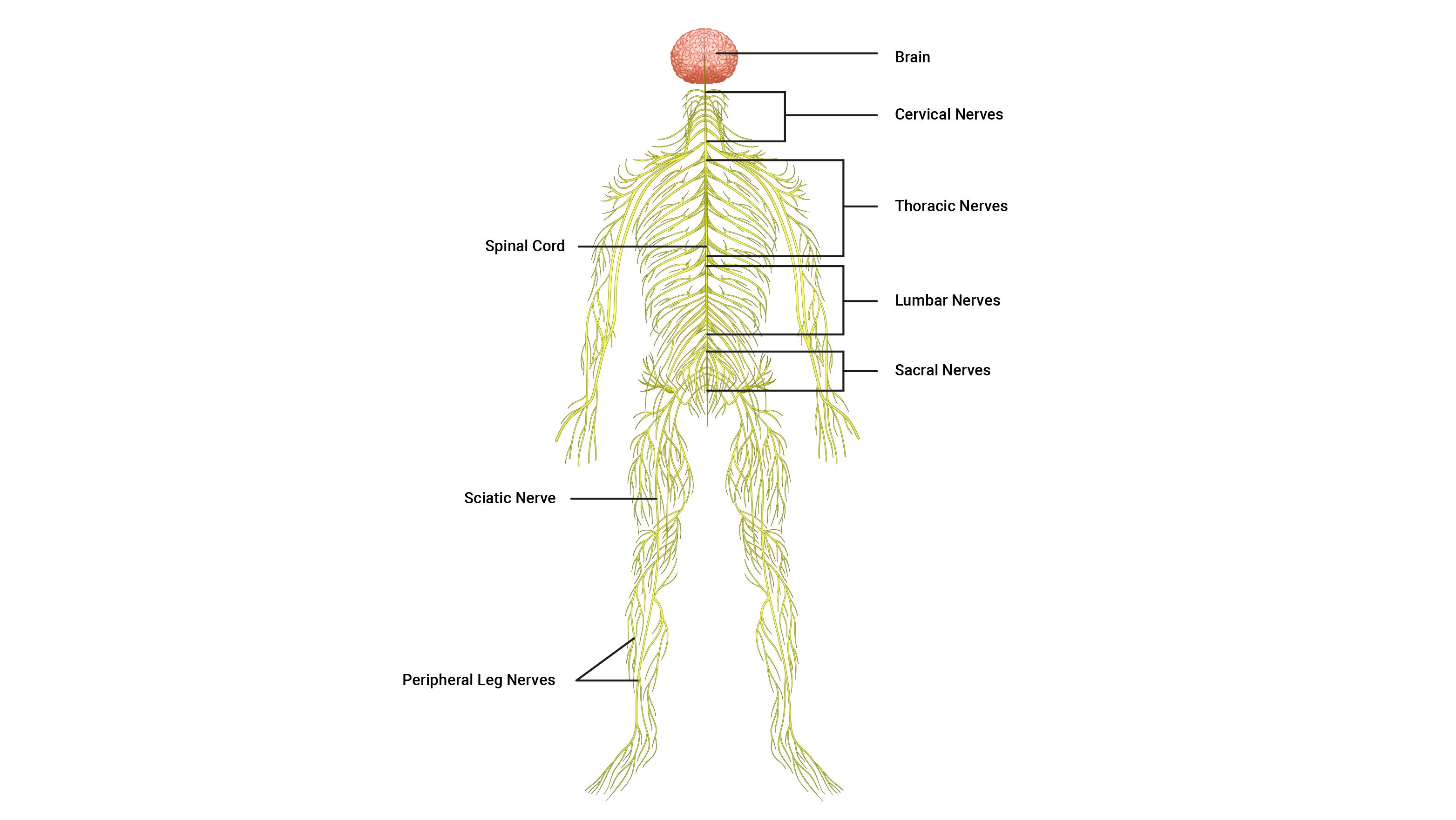
Hierarchical structure
The nervous system has many divisions, each with its own distinct purpose. Here you can see the hierarchy of the nervous system and how the various divisions are organised:

What makes up the nervous system?
While many components make up the nervous system, the 3 general classifications of components are:
- The brain: The brain is the supercomputer of the body. It processes billions of pieces of information and decides how and what we should focus on, respond to, or ignore.
- The spinal cord: The spinal cord is the superhighway for communication. It carries messages between the peripheral nerves and the brain. This also stores vital reflex commands for flight or fight responses.
- The neurons (nerves): Neurons are the communication pathways between the body parts and the spinal cord. They also carry impulses up the spinal cord and to and from the different parts of the brain.
Let's take a look at each component in closer detail, starting with the brain.
1. The brain
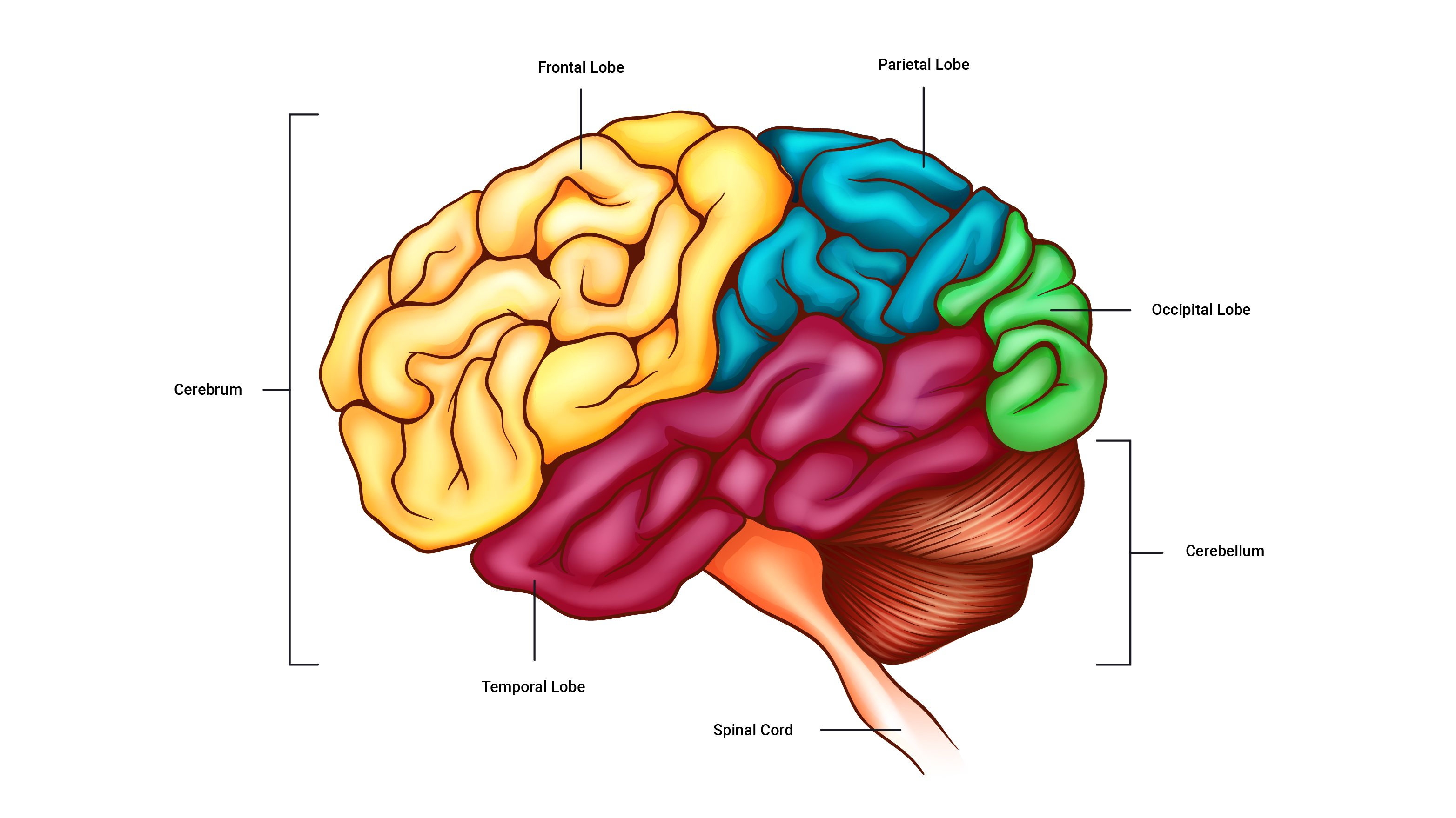
The brain is made up of about 85 billion neurons! They equip the brain with the communication tools it needs to perform its function. The adult brain is anatomically subdivided into 4 major parts:
- Cerebrum
- Diencephalon
- Cerebellum
- Brain stem
It is important to have a comfortable understanding of their general functions, so let's explore each part.
The cerebrum
The cerebrum is the largest part of the brain. It is comprised of 4 lobes responsible for different processes and actions. For example, thinking, movement control, sensory perception, and sight. What are the specific functions of each lobe? Expand the accordion to find out.
The diencephalon
The diencephalon is where we find the hypothalamus, as well as other parts which are responsible for controlling the body's internal environment, such as temperature control, regulation of heart rate and breathing, hunger, sleep and wake cycles and more.
The cerebellum
The cerebellum sits behind the brain stem. It coordinates movements and helps maintain body awareness.
The brain stem
The brain stem continues from the spinal cord and essentially connects the spinal cord to the brain. The brain stem has regulatory functions involved in breathing and heart rate.
2. The spinal cord
The spinal cord is a long, thin, incredibly fragile tubular structure encased within the spinal column and surrounded by cerebrospinal fluid. It is composed of nervous tissue and tracts of nerve fibres that allow two-way conduction of nerve impulses, both sensory and motor (input and output). This is a key piece of knowledge to remember.
3. Neurons
The third classification for the components that make up the nervous system is the neurons (nerves). Nerve anatomy is a large and complex area so we will look at this next.

Nerve Anatomy
Neurons are the individual pathways that carry electrical impulses around the human body. Nerves are bundles of neurons all heading in the same direction, with neurons branching off them at points to reach their intended destinations.
Functional classification of neurons
Neurons are responsible for producing complex networks and connections between:
- the brain and the spinal cord.
- the rest of the body and the brain and spinal cord.
These lines of communication are composed of highly specialised cells capable of transmitting highly complex information at great speeds. There are 3 main types of neurons. They tag team each other in the journey of signal transmission.
- Sensory neuron
- Interneuron
- Motor neuron

| Sensory neurons | Sensory neurons 'sense' information and provide the link between sensory receptors and the spinal cord. They transport electrical impulses to the spinal cord, where the impulses are passed to interneurons. |
|---|---|
| Interneurons | Interneurons are found only within the spinal cord and brain (CNS) and are situated between sensory and motor neurons. They process incoming sensory information received from sensory neurons and then activate the related motor neuron. They carry impulses up to the brain for processing and decision-making. 90% of all neurons in the body are interneurons. |
| Motor neurons | Motor neurons carry impulses that exit the spinal cord away from the CNS and are directed towards the effectors of the body, such as muscles, organs, and glands to cause action. |
Anatomy of a neuron
In reality, all neuron types look slightly different but contain the same key components. The picture below shows a generalised version of a neuron.
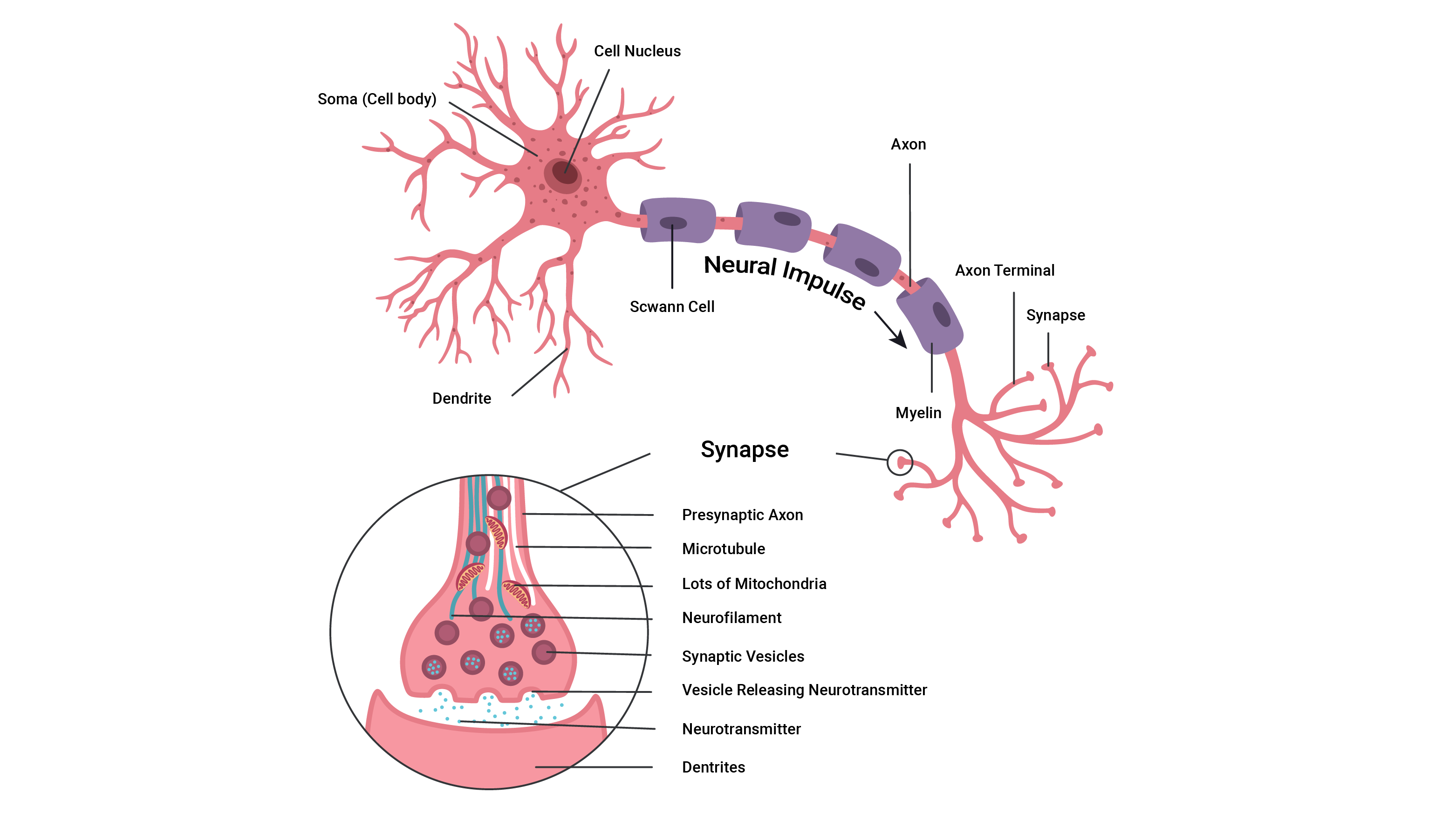
If you wish to learn more about the parts of a neuron, expand the accordion.
The control centre of the neuron is its cell body (also called the soma).
It has a centrally located nucleus which acts as the brain of the neuron.
The cell body usually gives rise to a few thick processes (sticky out bits) that branch into nerve fibres known as dendrites.
They house numerous sensory receptors for neurotransmitters and are the primary sites for receiving signals from other neurons.
The more dendrites a neuron has, the more information it can receive from other cells to incorporate into its decision-making.
All axons of the PNS (peripheral nervous system) have a sheath of Schwann cells around them. Each Schwann cell repeatedly spirals around the axon, putting down as many as 100 layers.
These layers form the myelin sheath. Think of the myelin sheath as similar to plastic around an electric cord. The myelin helps fully envelope the axon of the neuron to ensure there are no 'leaks' in information and to help speed the transmission of signals.
Myelinated nerve fibres conduct impulses faster than unmyelinated nerve fibres.
The axon is cylindrical and relatively un-branched for most of its length. It allows rapid transmission of nerve impulses from the cell body.
Axons may range from a few millimetres to more than a metre in length.
An axon typically connects with another muscle fibre or gland cell via its 'axon terminals'.
At the distal end, axons usually break into a complex network of fine branches. Each branch ends in a little swelling called a 'synaptic knob/bulb'.
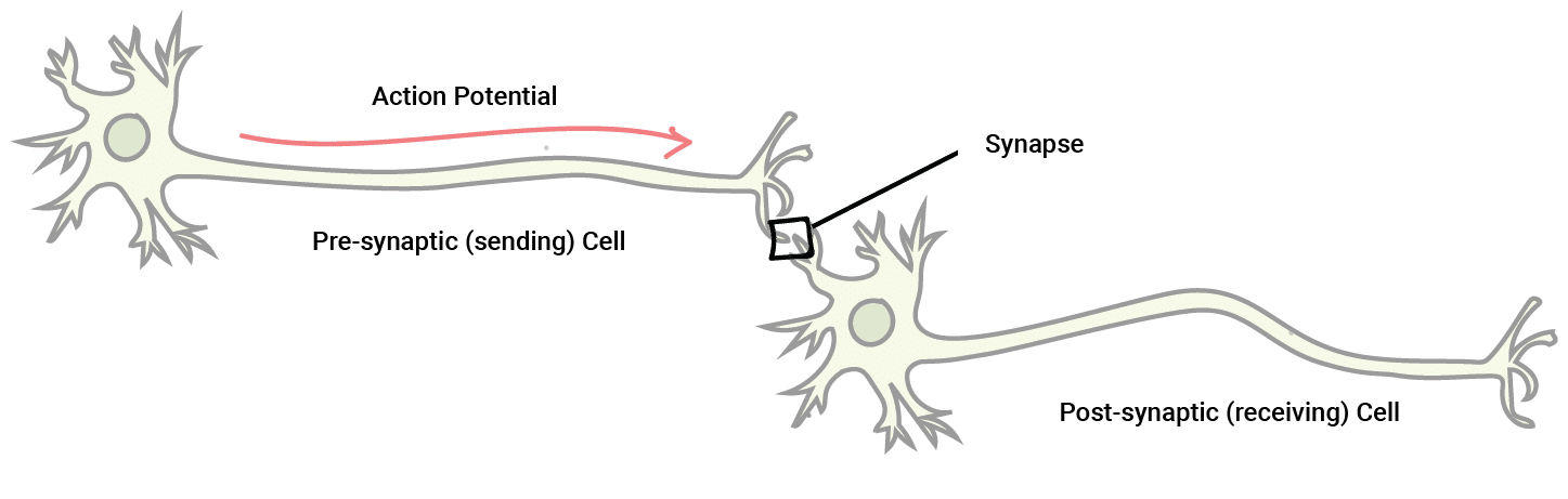
Each knob forms a junction with a muscle cell, gland cell, or another neuron. The point where communication occurs between two neurons or a neuron and an effector cell is called a Synapse.
Nerve Impulse
The currency of the nervous system is electrical impulses. These are also known as Action Potentials (AP). These electrical impulses have a voltage.
Nerve pathway
Almost every nerve impulse follows the same pathway, starting from a sensory receptor and ending at the effector muscles.
- Sensory receptors notice a change in the internal, or external, environment and send impulses via sensory neurons to the central nervous system (brain and spinal cord) for processing.
- Interneurons in the brain or spinal cord (CNS) process these impulses and decide to act, store the information, or discard it.
- If action is warranted, the CNS sends impulses down the spinal cord via interrnuerons, and out using motor neurons, to the body's effectors to cause action (for example, to the muscles, glands, organs).
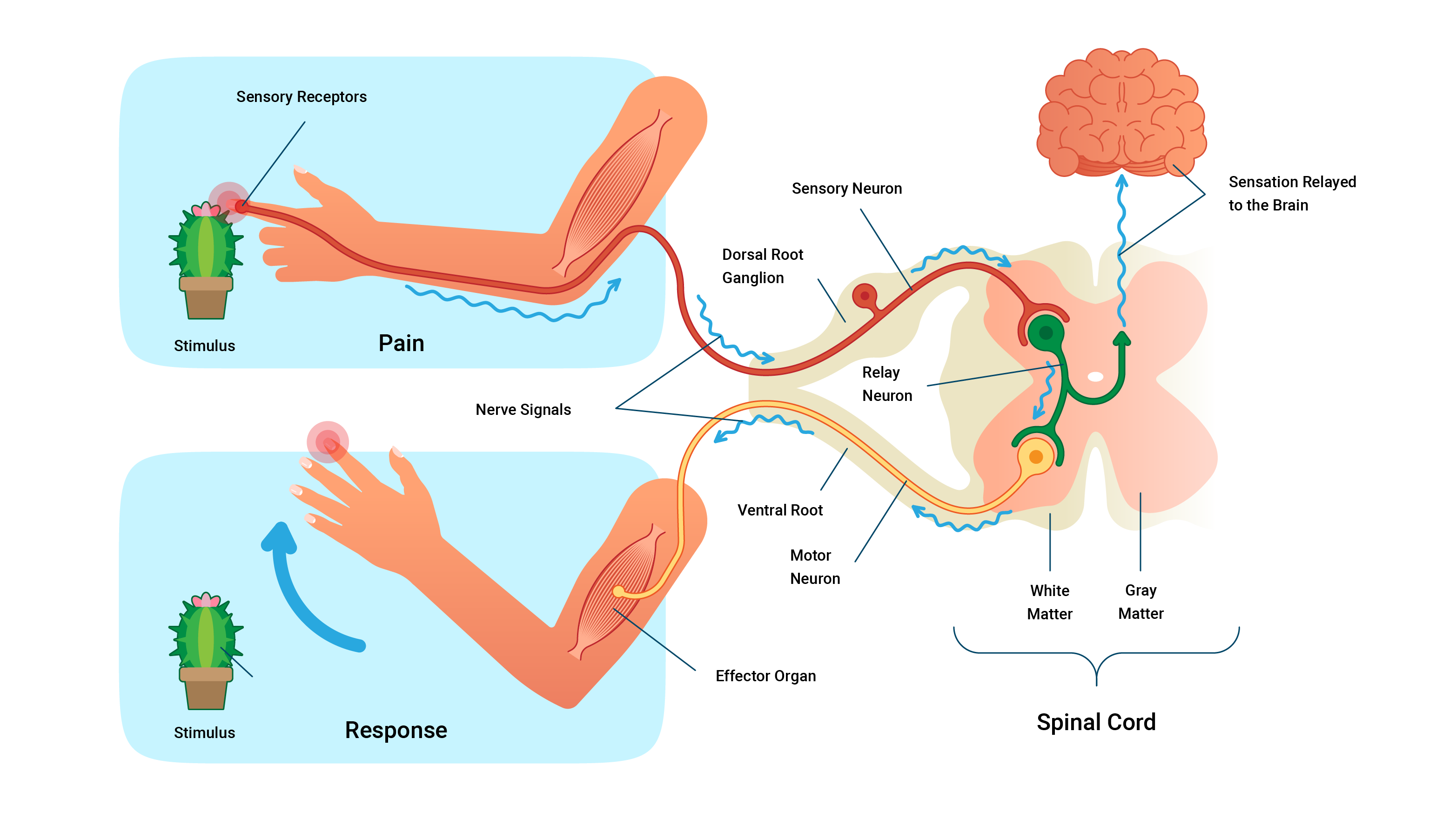
The image above shows the pathway of a nerve impulse. Sensory receptors notice pain from the stimulus (the cactus) and an impulse travels to the spine via sensory neurons. Once at the spine, interneurons travel up the spine to the brain where the sensation is relayed. The impulse travels back down the spinal cord via interneurons and out using motor neurons to the muscles where it activates the muscle fibres to perform a task - in this case, to move the hand away from the stimulus.
Check you have a good grasp of the nervous system before attempting your assessment and moving to the next topic.