Introduction video script:
(Visuals: athletes sprinting, cycling, and swimming, their breath visible in slow motion. A close-up of a deep breath being taken, lungs expanding.)
Whether you're pushing your limits in a race, powering through a tough workout, or simply recovering after exercise, there's one system working non-stop to fuel your every move—the respiratory system.
Your body’s cells continually use oxygen for metabolic reactions like nerve transmission, cell-to-cell communication, and muscle contraction. This means that oxygen O2 is always in high demand. Metabolic reactions create a waste product: Carbon dioxide (CO2). CO2 can become toxic to the body's cells when present at high levels. So, the respiratory systems ability to remove CO2 from the body is of equal importance to its role of supplying O2 to the cells.
In this module, we're diving into the incredible mechanics of how your body breathes, how it harnesses oxygen, and why this is critical for athletic performance. We’ll start by exploring the key structures—the lungs, diaphragm, and airways—and learn how they work in harmony to supply the oxygen your muscles crave.
Next, we will look at the physiology of breathing—breaking down how oxygen is absorbed into the body and delivered to tissues, along with how carbon dioxide is removed. Understanding this process is crucial for any athlete looking to maximize their performance and recovery.
Do you know how exercise impacts on the respiratory system? As an athlete, the way your body adapts to physical activity is key to improving endurance, power, and stamina. In this module, you'll uncover how the respiratory system responds to the demands of intense exercise and what happens to your breathing capacity and ability to exchange gases over time.
From the first breath of a newborn to the experienced endurance of an elite adult athlete, the respiratory system evolves across life stages. We'll explore how lung capacity, breathing efficiency, and respiratory health change from childhood, through adolescence, adulthood, and into older age. Understanding these changes will help you develop training programs that meet the needs of athletes at any stage of life.
"So, are you ready to explore the power behind every breath? Let’s get started!"
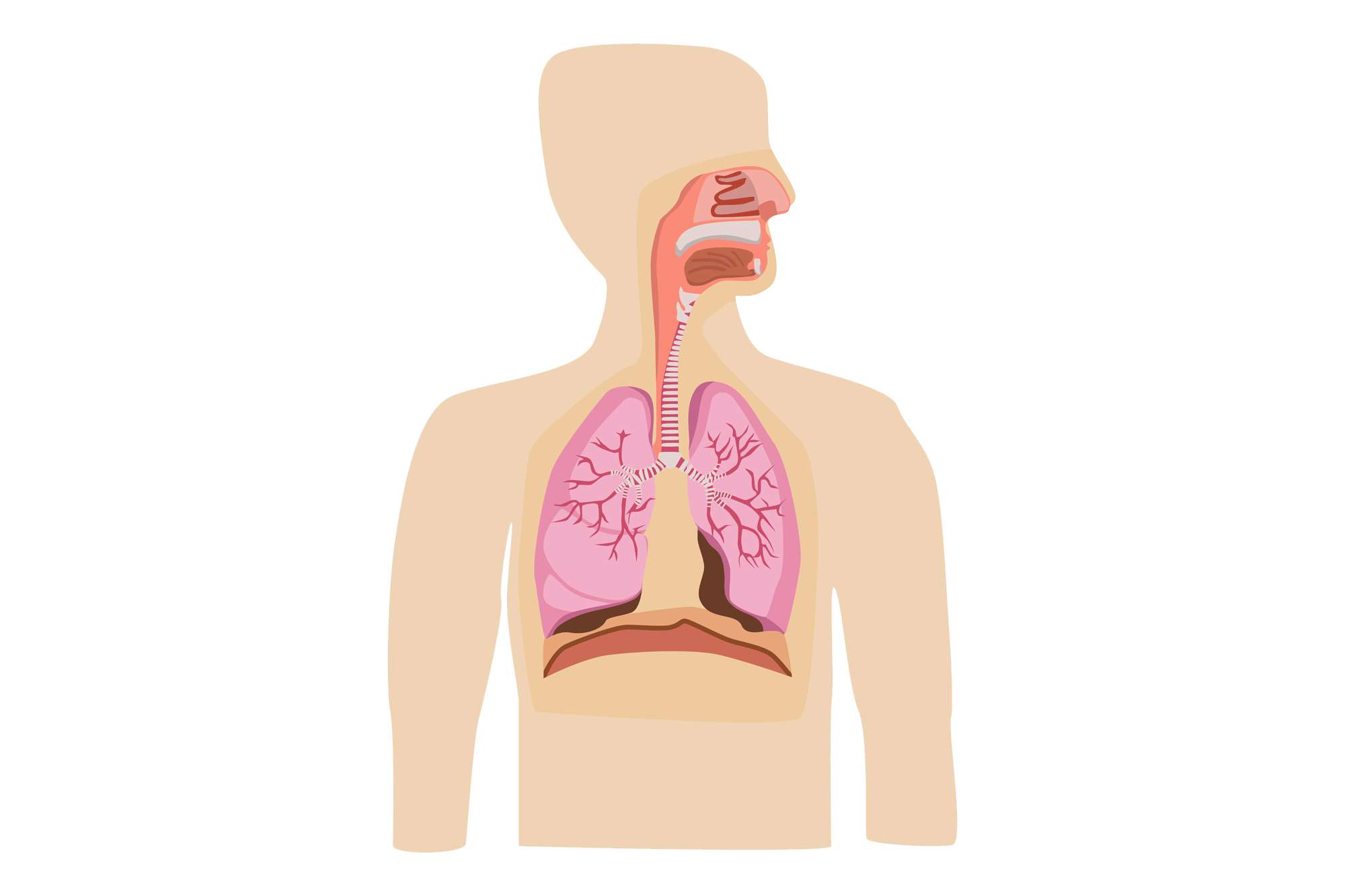
The respiratory system structures can be broken into two classifications:
The conducting division: These structures “conduct” the air into the lungs from the outside atmosphere. The conducting division includes structures including:
- The nose and mouth
- The pharynx (throat)
- The larynx (voice-box)
- The Trachea (windpipe)
- The Bronchial tree (larger and smaller tubes within the lungs)
Structures of the Conducting Division

They essentially provide passageways for the air to move through, however, they do a lot more than simply conduct the air. They also moisten, filter dust and debris from air and warm the air as it passes through.
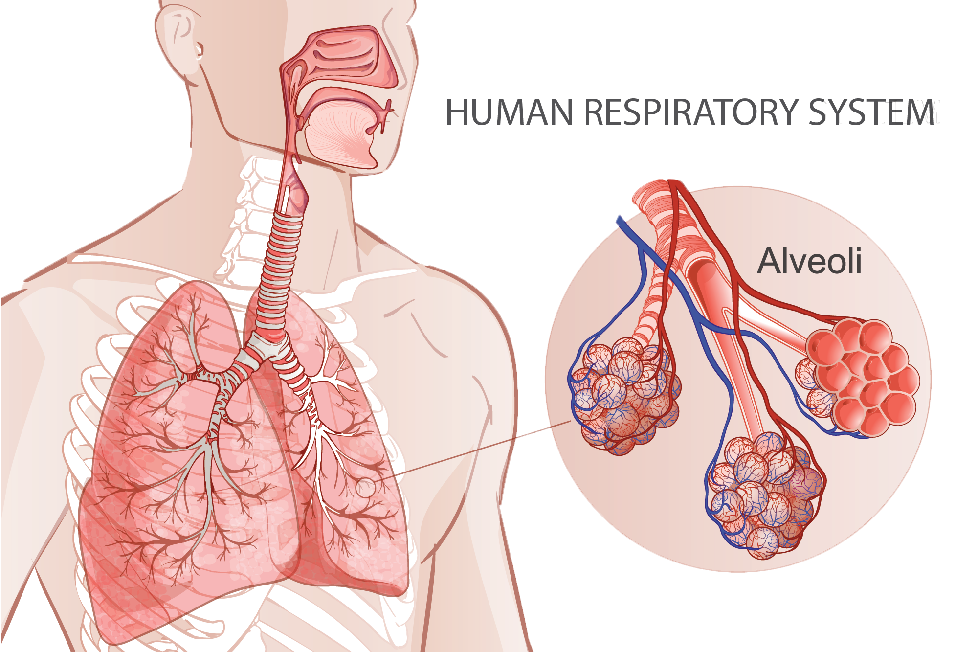
The respiratory division: These structures are directly involved in the gas exchange process (with blood). The main structures of the respiratory division are the alveoli (including their ducts and sacs).
Here is a quick overview of the respiration process and the role each structure plays in it. Watch the video, then read the information on each key structure of the system below.
Video Title: Respiration 3D Medical Animation
Watch Time: 1:43
Video Summary: This is a brief animation of ventilation and respiration. It covers the journey of air from the atmosphere to the lungs, and the transfer of oxygen from the alveoli to the red blood cells in the capillaries.
Post Watch Task: Watch the video, then read the additional information on each structure below.
Source: YouTube
Let’s take a look at each of the structures in a little more detail (including their key functions
The Nasal cavity
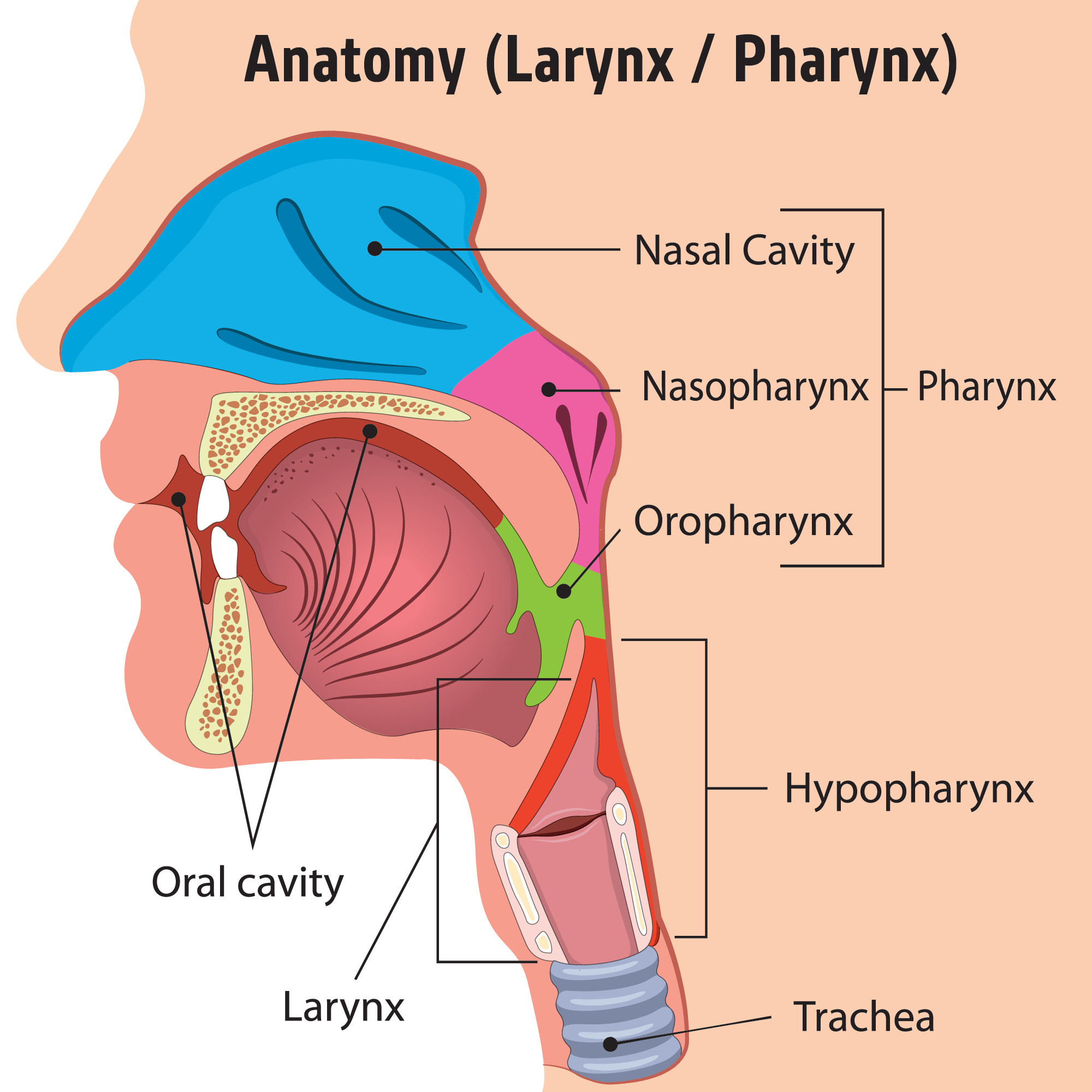
The nose and mouth are the entry points for air. The nasal cavity sits posterior to the nose and is the preferred method of inhaling air due to the functions it performs as air flows in.
These functions include:
- Warming the air: The lining of the nose and inside of the mouth are covered in tiny blood vessels in which travels warm blood. As air passes through the nose and mouth it is warmed. This is important for respiration as warm air inflates the lungs more easily than cool air.
- Humidifying the air: The nasal cavity contains a mucus producing layer that humidifies the air as it passes. This is important as humidifying the air allows it to reach the lungs more easily (instead as rising as warm air wants to do).
- Filtering the air – Tiny hairs within the nasal cavity trap dust particles, effectively cleansing the air for the lungs. The nasal cavity is structured so that air circulates multiple times as it passes through making maximum contact with these hairs.
- Protecting us from foreign invaders – The nasal cavity is the first line of defense (immunity). Tiny hair-like projections called cilia (along with mucus) help to keep harmful microbes from entering the body.
- Picking up odours – This alerts us to danger (and noxious smells that probably aren’t healthy to breath in).
Note: the mouth can also warm and humidify air but plays a lessor role in immunity and filtering of air. The mouth is typically used to inhale large quantities of air (like when we exercise). Ever tried to breathe through your nose while running? Try it sometime!
The Pharynx, Larynx and Trachea
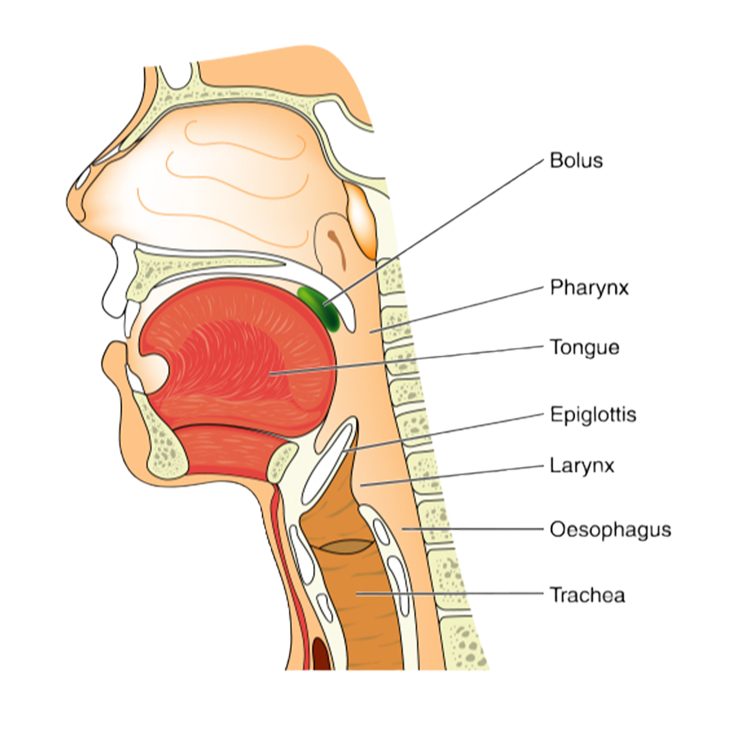
| Pharynx | The pharynx is part of the throat behind the mouth and nasal cavity, and above the oesophagus. It is lined with mucus-producing membranes that help you swallow. |
|---|---|
| Larynx | The larynx is also known as the voice box. It helps produce sound. It also contains the epiglottis, which closes off the windpipe when swallowing. |
| Trachea | The trachea is also known as the windpipe. It allows the passage of air and connects the larynx to the lungs. |
The Bronchial Tree
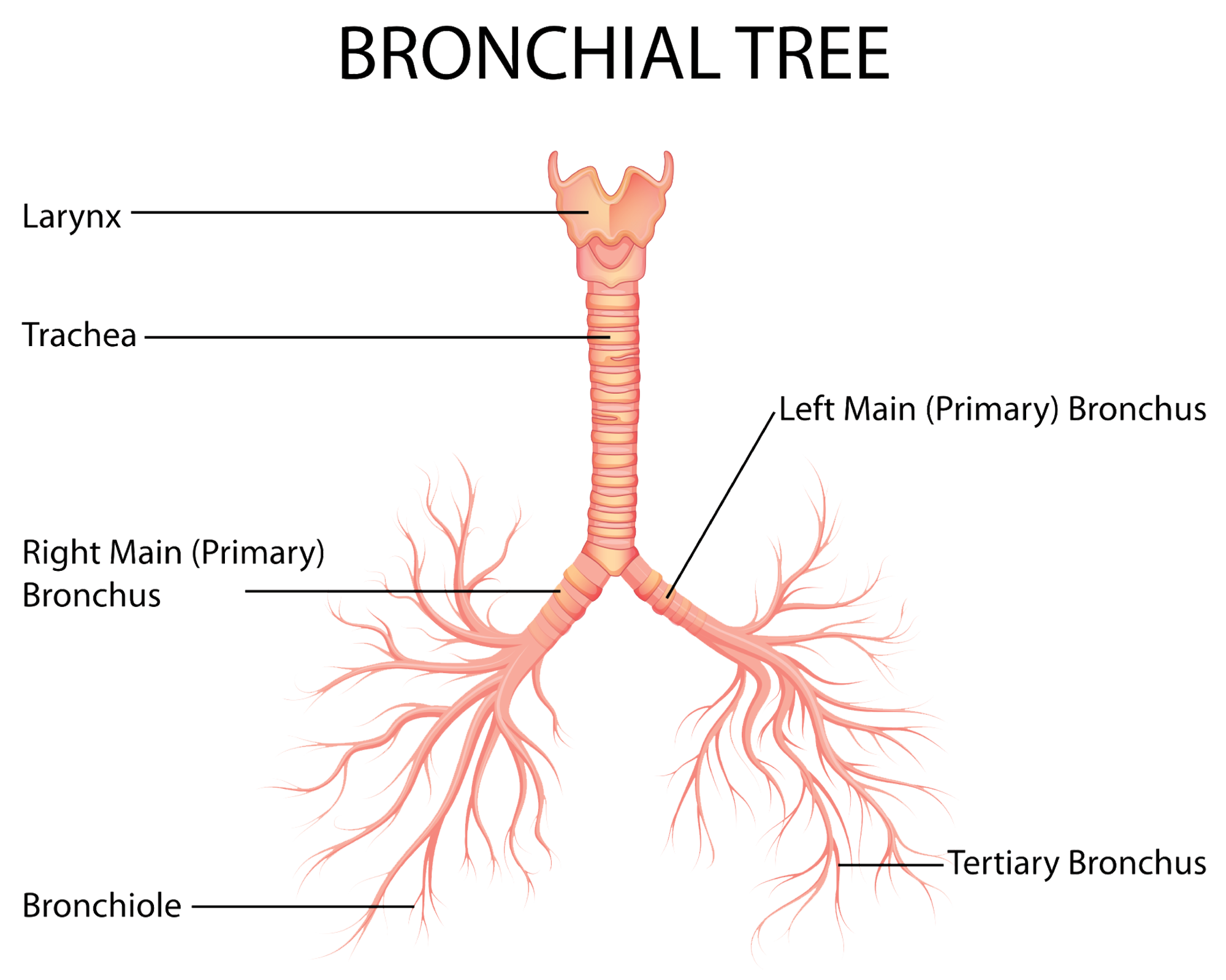
So named because it resembles an upside-down tree, with two large branches (Bronchial tubes or Bronchi) sprouting from the trunk (Trachea) to enter the left and right lungs, then smaller branches (the secondary and tertiary bronchi) passing the air deeper inside the lungs. The smallest “twigs” (Bronchioles) connect to the gas exchanging structures of the lungs (the Alveoli).
The Alveoli
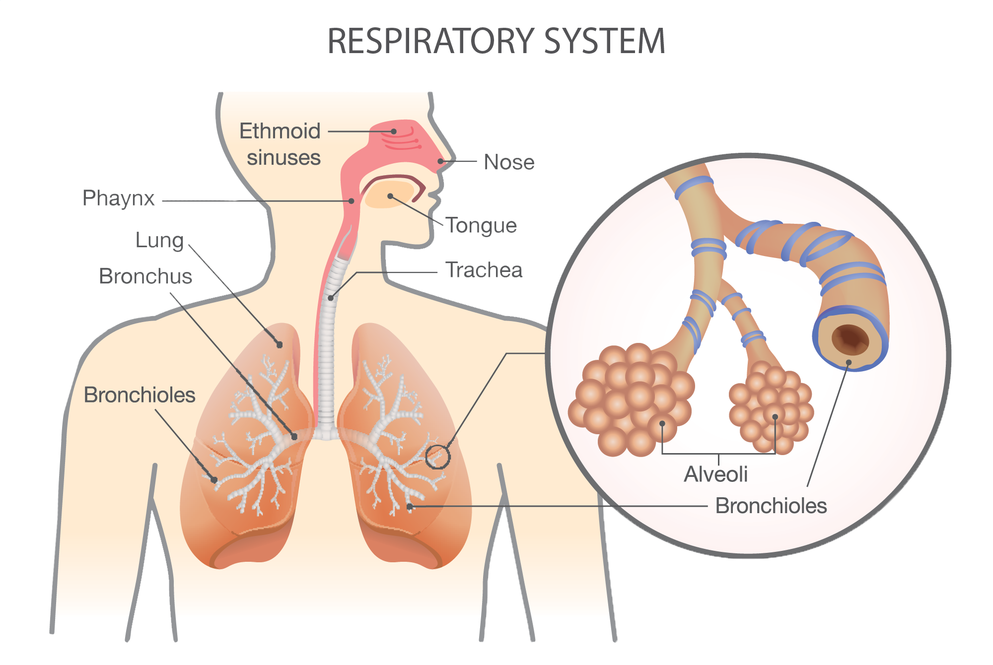
Alveoli are tiny air sacs at the end of the bronchioles. If the Bronchi are the branches, and the Bronchioles the twigs, then the Alveoli are the “leaves”. Just like the leaves of a tree take in CO2 from the atmosphere and produce oxygen, the alveoli take CO2 from the blood and supply the blood with O2. Eerily similar, don’t you think?
The Lungs
The lungs are cone-like structures. They are wider at the bottom where they rest on the diaphragm (a key respiratory muscle we will discuss soon), and narrower at the top where they form a point in the collarbone region. The flat outer sides of the lungs attach to the ribs (via membranes) and are referred to as the costal (rib) surfaces of the lungs.
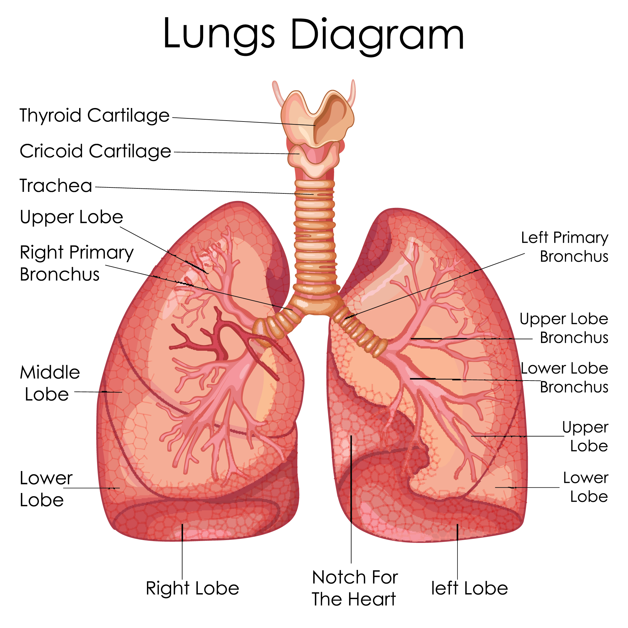
As you can see in the image, the left and right lung have some differences in their structure. The right lung (pictured on the left) has three lobes, while the left lung only has two. This is because the heart crowds into the left lung (which leaves it a “cardiac notch” to snuggle into).
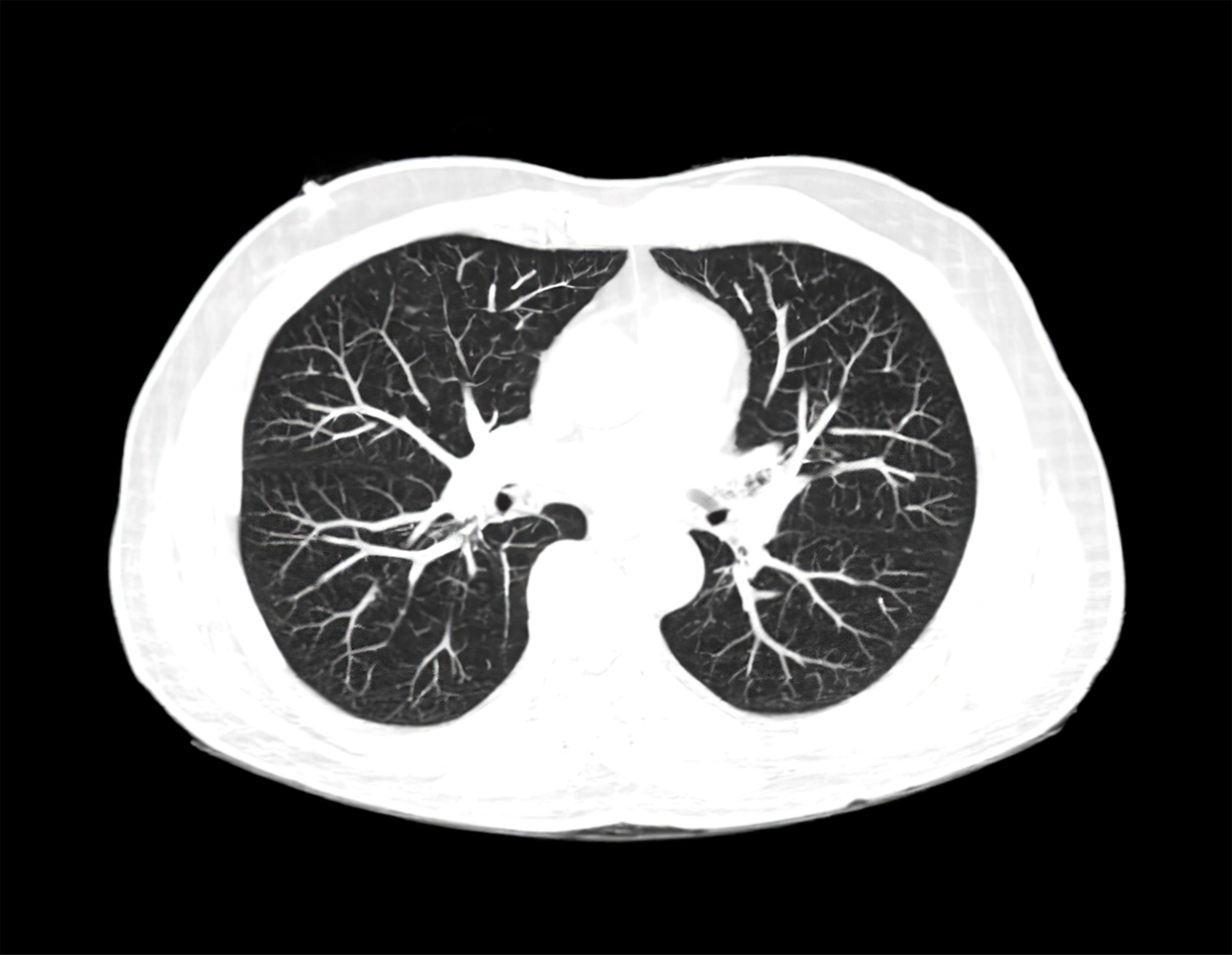
Contrary to popular belief, the lungs are not simply “balloons” of air. They are full of the structures of the bronchial tree that we have already discussed. Here is a superior view of an MRI of the chest that shows the lungs are anything but empty!
Recap of learning
The following series of questions are designed to check your retention of the content we have just covered on the structures of the respiratory system (and their basic functions).
The Respiratory System has one job and one job only. To bring O2 into the body and to remove CO2 from the body.
Your body’s cells continually use oxygen for metabolic reactions like nerve transmission, cell-to-cell communication, and muscle contraction. This means that oxygen O2 is always in high demand.
Metabolic reactions create a waste product: Carbon dioxide (CO2). CO2 can become toxic to the body's cells when present at high levels. So, the respiratory systems ability to remove CO2 from the body is of equal importance to its role of supplying O2 to the cells.
The process of getting air in and out of our lungs may seem like a simple process, but it is actually a well-orchestrated sequence of muscle action, changes to lung size and manipulation of pressure that is all governed by the autonomic nervous system. To understand the process of breathing, you need to have an awareness of the key muscles we use to manipulate the lungs, along with knowledge of the principles of air flow.
This task is designed to check your understanding of this content following your face last face to face lesson. Feel free to read over the notes related to breathing mechanics in your learner resource before attempting these questions.
There are a number of measures that can be used to predict respiratory efficiency. Some require invasive procedures or expensive equipment to calculate, while others are fairly simple measures. The following questions are designed to check you understanding of the common respiratory measures we covered in our face-to face-learning. It will also assess your knowledge of the way the initiating exercise affects some of these measures.
We now know exactly how air makes its way in and out of the lungs.
The final part of the respiration process (and the reason we breath), is to exchange gases with the blood, so it can deliver oxygen to the cells and off-load CO2.
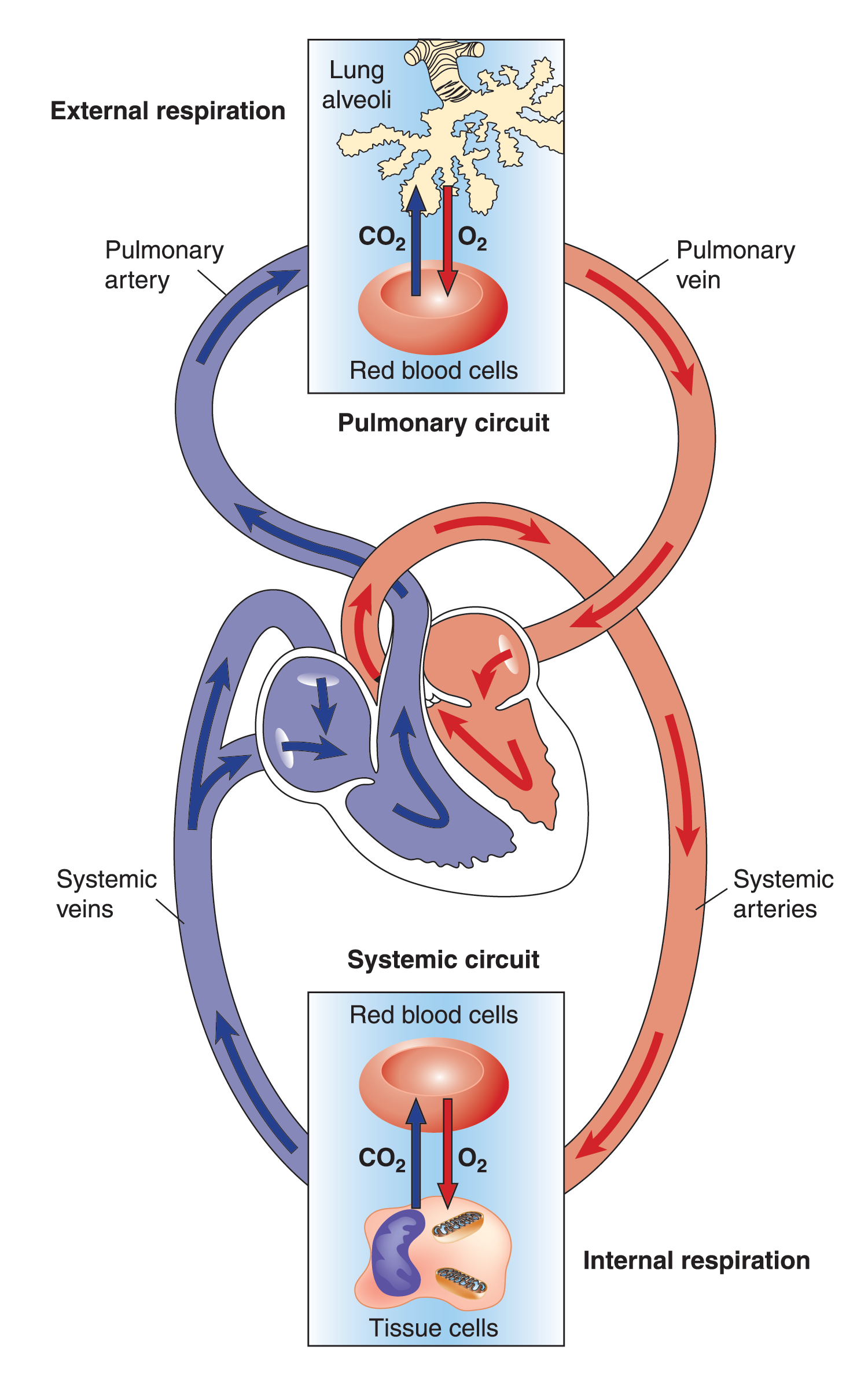
There are two points for gas exchange in the body:
- Between the lungs (alveoli) and the blood (hemoglobin)
- Between the blood (hemoglobin) and the tissues (cells)
Diffusion
Before we can discuss gas exchange in detail, there are a couple of things you need to know.
“Diffusion” is the term that describes the movement of gases from one area of the body to another. In this instance, diffusion occurs through very thin membranes (like the walls of capillaries).
The rule of gas diffusion is the same as the rule of air flow, in that:
Gases will only diffuse from an area of high gas pressure to an area of lower gas pressure
The Pressure of Gases (PO2)
Air is made up of a combination of gasses. The largest proportion of air is Nitrogen (78%), followed by Oxygen (21%).
The composition of air
Each gas in a mixture exerts a portion of the total pressure of the gasses combined. Air (at sea level) has a total pressure of 760mmHg. If Oxygen is 21% of air, then the pressure of Oxygen (PO2) is 21% of the total air pressure (i.e., 0.21 x 760mmHg = 159mmHg).
The more of a gas that is present in an area, the more pressure the gas exerts. For example, an area that is high in O2 would have a high PO2, whereas an area that is low in O2 would have a low PO2. The same is true for areas containing CO2.
The rule of gas exchange states that gases diffuse from high to low pressure. This can be simplified to say that gases diffuse from an area of high concentration (of the gas) to an area of low concentration (of that gas) until the concentrations of the gas are even.
Put simply, if one part of the body has lots of O2 and comes into contact with an area of the body that has low levels of O2, then O2 will diffuse into the area of low O2 until the concentrations become even in both areas. The same is true for CO2.
Here is an example to help you understand.
 |
 |
|
A jar with a divider through the middle has pure oxygen pumped into one end and pure CO2 pumped into the other. |
|
Pulmonary Diffusion
The first point of gas exchange is in the lungs and occurs between the alveoli and the pulmonary capillaries. The lungs take in fresh air and conduct this air (rich in O2) to the alveoli. Therefore, the pressure of O2 (PO2) in the alveoli is high (because there is lots of O2 present).
The blood returning to the lungs is lower in O2 because some has been consumed by the body tissues. Therefore, the PO2 in the pulmonary capillaries is low.
When these two structures come into contact, the pressure difference between the two areas drives O2 molecules through the alveolar membrane and capillary wall and into the bloodstream (oxygenating the blood). This means that the blood that heads back to the heart (and then is pumped out into the body) now has a high PO2.
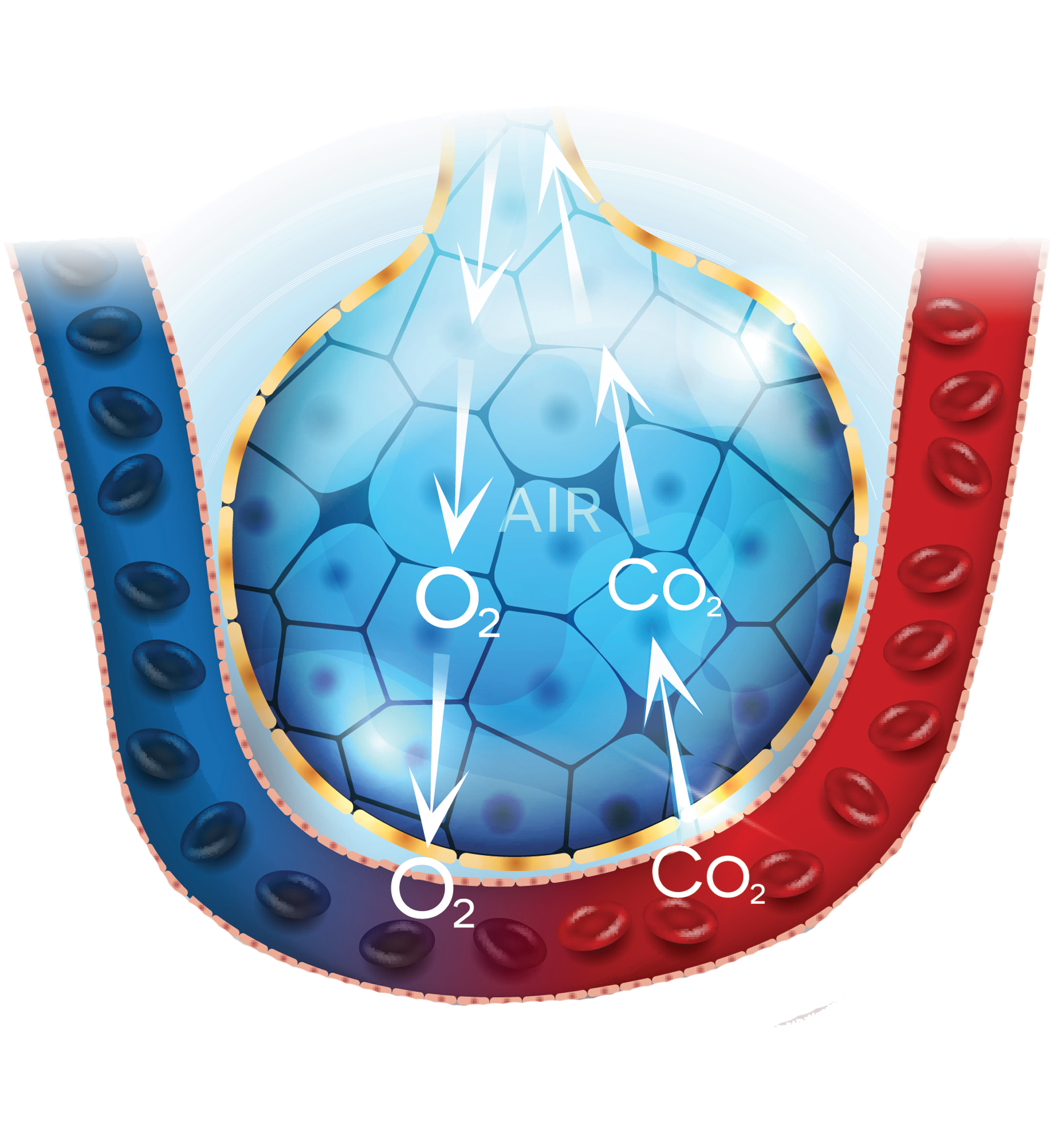
Conversely, the CO2 levels (and therefore PCO2) in blood arriving back to the lungs (from the body) are elevated because the cells have produced it. The CO2 levels in the alveoli are comparatively lower, as CO2 is breathed out of the lungs in each breath cycle. When the pulmonary capillaries come into contact with the alveoli, the difference in pressure of CO2 (PCO2) drives CO2 through the capillary wall and alveolar membrane into the alveoli for removal. This exchange can be seen on the image below. Blood heading back to the heart (and out to the body is now low in CO2).
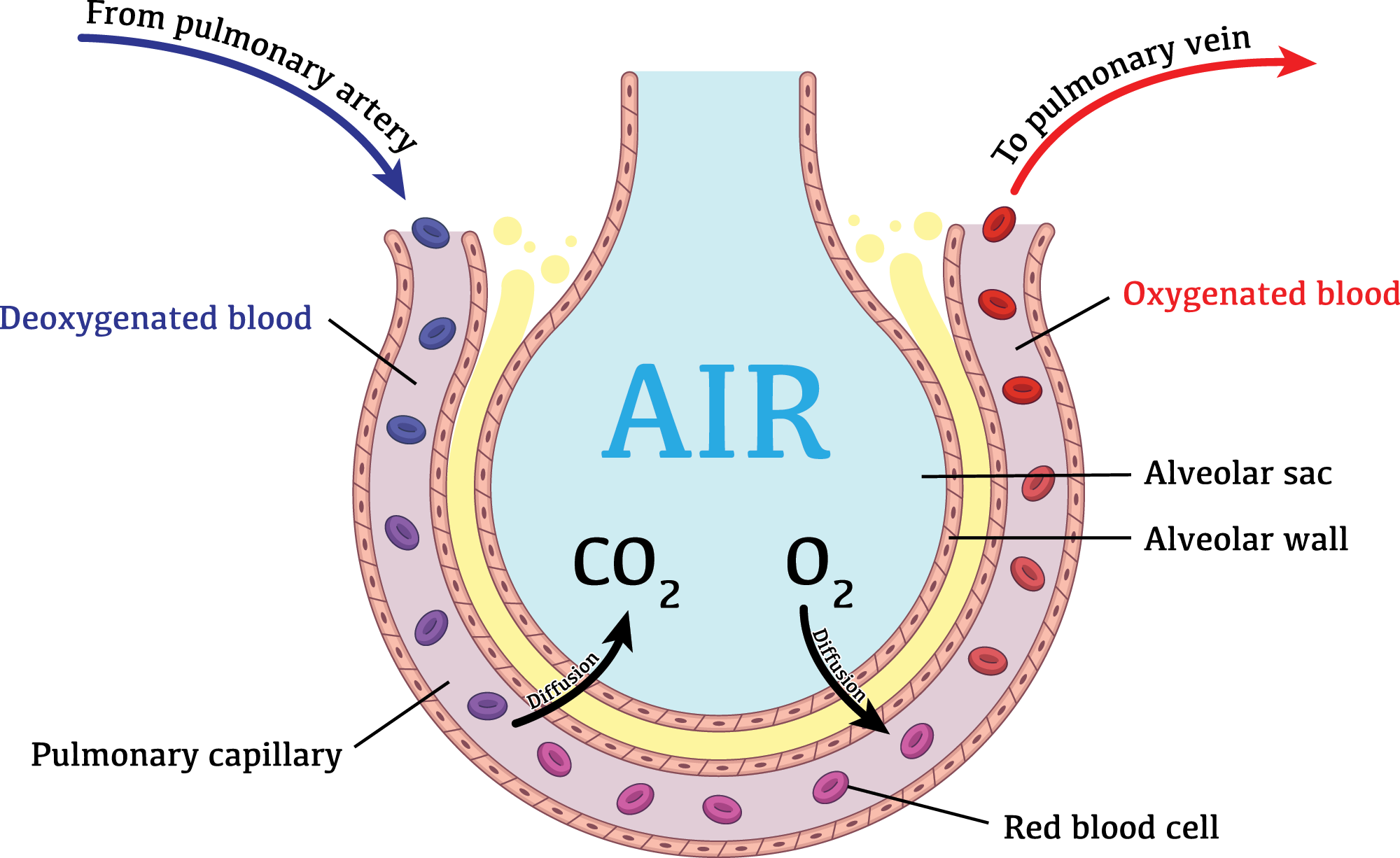
This short video gives a visual overview of the process of pulmonary diffusion. Watch the video, then complete the task that follows.
Video Title: Gaseous exchange in the lungs
Watch Time: 1:52
Video Summary: This video explains how gas exchange occurs in the lungs between alveoli and pulmonary capillaries.
Post watch task: Watch the video, then complete the task that follows.
Source: YouTube
Show your understanding of the pulmonary diffusion process by dropping the words into the appropriate place on the image. Remember the terms PO2 and PCO2 simply reflect how much of the gas is present in that area, e.g., high means lots, low means not much.
How Oxygen is Transported in blood
Haemoglobin (Hb), present in red blood cells, carries about 98.5% of O2 within blood (one haemoglobin molecule can carry four O2). The remaining 1.5% of O2 is dissolved in blood plasma.
The O2 carrying capacity of untrained males and females is:
- 201 mL O2/L blood in males
- 174 mL O2/L blood in females
Myoglobin (Mb) is an iron/oxygen-binding protein found in muscle cells that shuttles O2 from the cell membrane to the mitochondria within the muscle cell. Mb has a higher affinity (binding desire) for O2 than haemoglobin, which allows Mb to store a limited amount of O2 'on-site'.
It doesn't take long for haemoglobin to become fully saturated (i.e., carry four O2 per molecule). In times of increased heart rate (e.g., exertion, fright, or illness), red blood cells spend less time at the alveoli, (as little as 0.3 secs), but there is still enough time for haemoglobin to become fully saturated with O2.
This great video explains the role that red blood cells (and haemoglobin) play in the transport of oxygen. It also briefly explains how red blood cells process CO2 at the tissues and deliver it back to the lungs. Watch the video then answer the short series of questions that follows.
Video Title: How the oxygen you breathe gets delivered to the cells of your body.
Watch Time: 2:18
Video Summary: This video explains how oxygen is carried in blood and exchanges with the tissues. It also explains how carbon dioxide is transported back to the lungs for removal.
Post watch task: Watch the video, then complete the task that follows.
Source: YouTube
Check your understanding of the oxygen and carbon dioxide transport process in blood by answering the short series of questions below.
H5P here
Diffusion of gases at the cells (Internal Respiration)
Now that you understand the principle of gas exchange, the diffusion of gasses at the tissues should be easy to understand. Arterial blood is now carrying oxygen rich blood to the body cells, so they can use it to create energy. This means the blood arriving at the cells has a high PO2.
The body cells have a lower PO2 as they are constantly using oxygen. The more a cell is working, the lower the PO2 will be in that cell. For example, a muscle fibre that is contracting will be consuming more oxygen than a muscle fibre that is resting. The larger the difference in PO2 between the blood arriving in systemic capillaries and the cell, the more oxygen that will be driven into the cell.
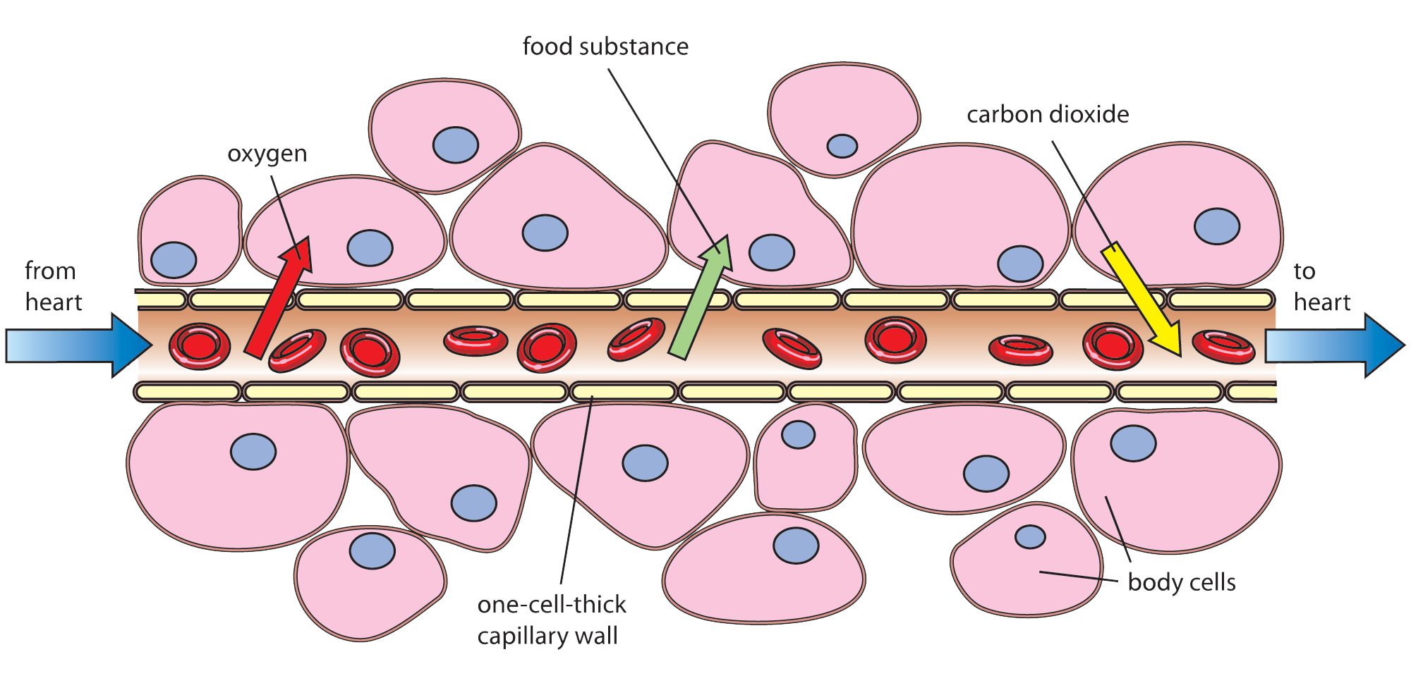
As CO2 is produced by the cells, the PCO2 levels in the cell are higher than in the blood arriving. This forces CO2 to leave the cell and enter the bloodstream (as shown in the video above). The more active a cell is, the more CO2 it will be producing and the higher the PCO2 difference will be forcing greater amounts of CO2 into the blood stream. This raise in CO2 in the blood is what signals the brain to increase ventilation rates.
This short video gives a visual demonstration of internal respiration (gas exchange at the cells). Watch the video, then show your understanding by completing the short task that follows.
Video Title: Internal Respiration. How oxygen and carbon dioxide are exchanges at the cells
Watch Time: 1:15
Video Summary: This video explains how oxygen is exchanged carbon dioxide at the cells.
Post watch task: Watch the video, then complete the short task that follows.
Source: YouTube
Show your understanding of the internal respiration process by dropping the words into the appropriate place on the image. Remember the terms PO2 and PCO2 simply reflect how much of the gas is present in that area, e.g., high means lots, low means not much.
Regular exercise elicits favourable adaptions across most organ systems in the body. The respiratory system is no exception. One of the most noticeable effects of getting fit, is the ability to breathe easier during exercise. This is due to a series of changes that the body makes due to the repeated stress of exercise. When a body is regularly put in a position where there is increased demand for oxygen delivery to muscles and CO2 removal, it adapts to make the process easier.
The following questions are designed to check your awareness of the chronic effects of exercise on the respiratory system.
H5P here
End of Topic
That’s it for the respiratory system. You have now completed the next 3 topics of the module! Now that you have completed the content for the muscular, cardiovascular and respiratory systems, you are ready to prepare for the second of your assessment events for this module.
Your second assessment event for this module is scheduled for your next face-to-face lesson.
Assessment 1 (Part B) is an open book assessment conducted under test conditions. It will assess the last three topics that we have covered in your learning – The muscular, cardiovascular and respiratory systems.
The assessment is made up of ten questions relating to three different case study topics. The case study scenarios cover the following topics:
- Muscle terms
- Muscle changes across the life stages
- Chronic effects of exercise in muscle
- Muscle imbalance and correction
- Chronic effects of exercise on the cardiovascular and respiratory systems
- Using heart rate training zones
You will be able to bring your learner resource and any other written notes into class.
Make sure, if you have missed any classes over the last 3 weeks, that you fill in any of the missing exercises in your learner resource. You can access any of the class presentations in your class teams under Module 2, weeks 3 to 5 to help you do this.
This final segment of online learning is to make sure you are fully prepared for the assessment. To complete the tasks below, you will want to have your learner resource handy, along with any other notes you have taken across the last 3 weeks of learning.
The following tasks will present case study scenarios that are similar too, but not the same as those you will be tasked with in the assessment. The assessments for this topic are not simply information recall. The questions are designed to assess your ability to apply what you have learned to the case study scenario presented.
What you need to do
The answers to these tasks will require you to find the relevant information that you need in your learner resource, then write it up as written paragraph answer (kind of like what you need to do in the assessment!).
Each case study scenario introduced will provide the topic knowledge that the answer will require. This way you can turn to the relevant section of your learner resource for easy access to the information you need.
Scenario 1
(Topic – Muscle terms)
A new PT client is wondering about the different types of exercise that have been included in their program. They are curious about why some focus more on the lowering of weight (slowly and controlled), while others focus more on lifting the weight up, or holding the weight in place for a number of seconds. Explain the three main types of contraction used in resistance training and why it is important to focus on all three during sessions.
H5P Essay
Scenario 2
(Topic – Agonist and Antagonist Relationship)
One of your classmates is confused about the different roles that muscles play during exercises. They know that some muscles are the main movers and that some help with a movement, but they don’t understand why other muscles would resist a movement?
H5P Essay
Scenario 3
(Topic – Types of exercises )
Your friend shows you a program they have written themselves for the gym. Here is a list of the exercises they have chosen:
- Bicep curls
- Triceps extensions
- Lateral raises
- Front raises
- Rear delt flies
- Leg extensions
- Hamstring curls
- Calf raises
You ask your friend what her goal for the program is and she tells you it is weight loss.
H5P Essay
Scenario 4
(Topic – Gender Differences in Muscle Development)
Your 12-year-old niece is a talented rugby player. Throughout primary and intermediate school, she has always played rugby with a mainly boys’ team. This year, she has been told that (for her own safety) she must play in an all-girls team. She doesn’t understand why she can’t keep playing with the team she has been playing with the last few years.
H5P Essay
Scenario 5
(Topic – Muscle Fibre Types)
A friend of yours has heard that unless you have the right genetics, there is no point in even training for a sport.
H5P Essay
Scenario 6
(Topic – Muscle imbalance)
You notice a client at the gym is having trouble with maintaining good posture in a few exercises. When you observe her standing during an exercise break, it is clear that she has some form of muscle imbalance. Her upper back is quite rounded, her shoulders roll forward and her neck sits forward in standing. You strike up a conversation with her and find out that she is a computer programmer. You think you might be able to help her with this issue.
H5P Essay
Scenario 7
(Topic – Muscle and Aging)
Your grandfather complains that he is having trouble getting out of his favourite armchair. He says that lately, he just doesn’t seem to have the strength in his legs to push up, so has to rely more on his arms to push himself up to standing. He is worried that something is wrong with him.
H5P Essay
Scenario 8
(Topic – Chronic effects of Exercise on the CV & Respiratory Systems)
6 weeks ago, you began swim training with the aim of completing your first ocean swim race this summer. When you first started training , you found it really difficult. You had trouble regulating your breathing and could only manage a couple of lengths before having to stop and rest. You are now finding things much easier! In your last session you managed 40 laps without stopping!
H5P Essay
Final practice task
This task is designed for you to practice your ability to use the Karvonen formula for working out target heart rate training zones for different clients. The following flash cards have information relating to different case study clients. Use the information on the card (and the Karvonen formula) to work out the target heart training zone asked for on the card. Once you think you have the answer, type it in the space at the bottom of the card then check your answer to see if you are correct. Answers should be whole numbers only (round up or down) and don’t need a measurement value (e.g. bpm).
Note: If a range is asked for (e.g., 60-65%), work out the 60% figure, then the 65% figure and write them both separated only by a ‘-‘ (dash – no spaces), e.g., 138-145
