In this topic, we focus on the structure and function of the skeletal system. You will learn:
- the different functions and shapes of bones
- how to identify types and location of bones
- the structure and function of the spine
- the effect of exercise on bone density
- body cavities, planes and sections
- movement types and directional terms.
Terminology and vocabulary reference guide
As an allied health professional, you need to be familiar with terms associated with basic exercise principles and use the terms correctly (and confidently) with clients, your colleagues, and other allied health professionals. You will be introduced to many terms and definitions. Add any unfamiliar terms to your own vocabulary reference guide.
Activities
There are activities throughout the topic and an end of topic automated quiz. These are not part of your assessment but will provide practical experience that will help you in your work and help you prepare for your formal assessment.
Welcome to your very first system of the human body, the skeletal system!
This is one of eleven systems in the human body. It takes eleven systems to work both independently of each other, in interchanging pairs (of two or more) to maintain our daily health and functions (homeostasis,which is maintaining our internal and external balance)
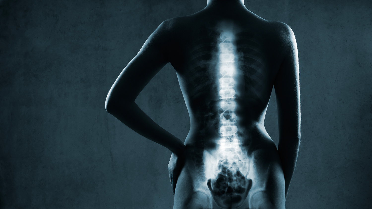
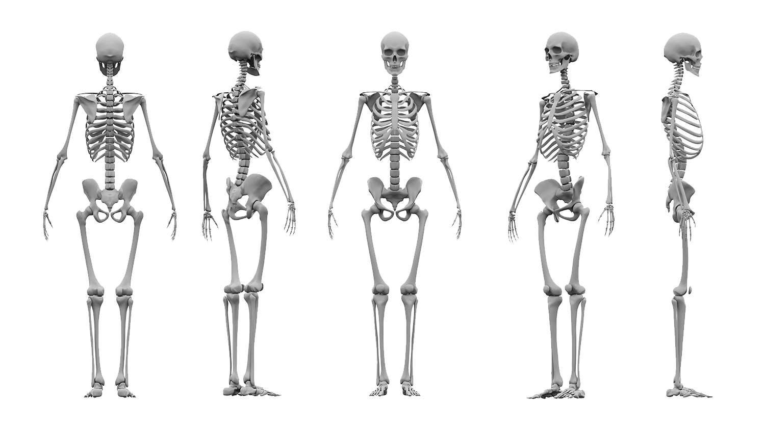
In this topic, we will take a closer look at bones, what they do, how they grow, and how they all fit together to make up the skeleton (in other words, their 'structure and function'). The images below show the skeleton from different perspectives.
Bone development
Did you know?
Bone is composed of several types of tissues working together. These are bone, cartilage, adipose, connective tissue, and nervous tissue; this is why each individual bone in your body is technically considered to be an organ.
Bone is classified as a dynamic living tissue as it continues to perform a process called 'remodeling', which is the process of new bone-forming and the breaking down of old bone. Pretty cool right?
Bones determine your body's dimensions, that is, your size and proportion. Our bones begin to form after just six weeks as a foetus and continue to grow until a person is approximately 25 years of age.
Research suggests peak bone massage may be considerably lower for females. Scientists think it is now important for young women to endeavour to build 'bone banks' while young to lay down as much bone as possible before they turn 20 years old. To do this we must ensure adequate calcium in our diet and perform regular loaded exercise.
Functions
The skeletal system holds the responsibility for six main functions.
- Support
- Protection
- Movement (assistance in movement)
- Blood cell production
- Mineral storage and release
- Storage (adipose/fat storage).
It is important in your fitness career to become familiar with these as this will support you in achieving a more complete understanding of the human body as well as medical conditions that affect the skeletal system. It will also help you effectively plan an exercise program around these.
Support
The skeleton provides a structural framework, like scaffolding, which provides a structure for our body (otherwise, we would essentially be a 'blob') as well as providing an attachment site for muscles, ligaments, and tendons.
Protection
Bone is the hardest substance in your body. It is used in many places to protect the vulnerable organs underneath from outside forces. Examples include the ribs protecting the vital internal organs such as the heart and lungs and the cranium protecting the brain. Remember, structure mirrors function; there is a reason why nature designed us so!
Movement (assistance in movement)
Bones provide attachment points for ligaments and tendons. Muscles are attached to bones via tendons. The bones act as rigid levers for movement to occur. This movement is articulated by joints (where two or more bones meet and are joined by ligaments). When a muscle contracts, it shortens and pulls on the bone it is attached to, as a result, movement occurs.
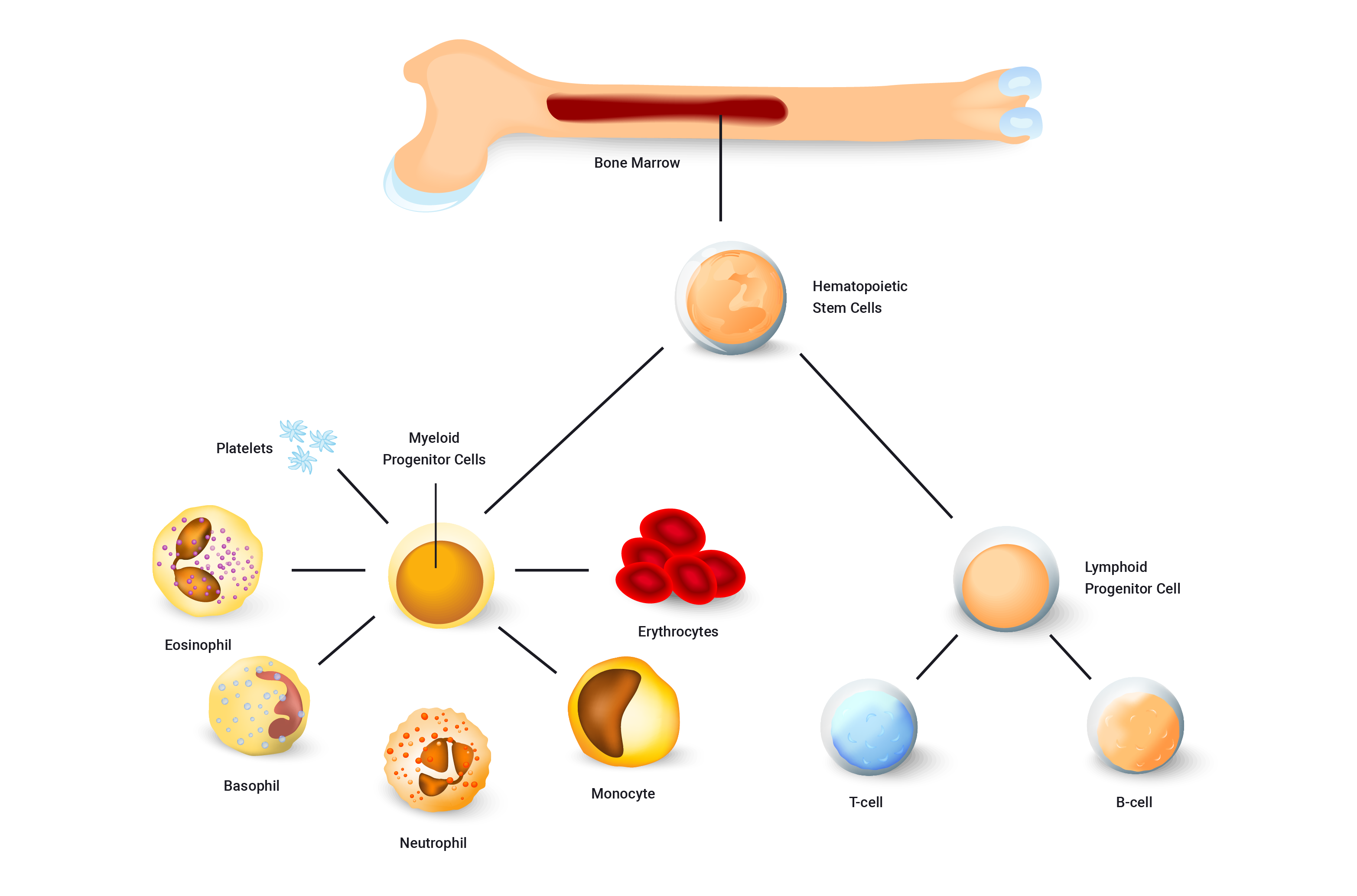
Blood cell production
Both red and white blood cells are formed in the bone marrow. The previous picture shows the many blood components that are produced in the bone marrow.
Mineral storage and release
Bone is the body’s main storage of calcium, it stores approximately 99% of the body’s calcium and releases when needed. Calcium is hugely important for bone growth and development as well as for many other cells in the human body (to communicate with each other and aid in functions such as muscle contraction).
Storage (adipose/fat storage)
Fat tissue is also stored within the bone in the bone marrow, which is additional energy storage and can be released when required to maintain physiological processes.
Watch the following video which provides an overview of the major structures of bone.
Shapes
Bones come in different shapes and sizes. The shape and size depend on the role of the bone. Structure once again mirrors function.
There are five different shapes of bone, these are.
- flat bones
- long bones
- short bones
- irregular bones
- sesamoid bones.

Let's take a closer look at these and their structure and function.
Flat bones
Flat bones offer protection and many sites for ligament and tendon attachment. They provide coverage of large areas. Examples of these bones include the:
- cranium
- ribs
- sternum
- scapula
- pelvis

Long bones
A long bone is one that is typically longer than wide. Long bones can also act as levers for muscles to act on. Examples of these bone include the:
- humerus
- radius
- ulna
- femur
- tibia
- fibula
- metacarpals and metatarsals
- phalanges.
A typical long bone consists of the parts/regions shown in the following diagramme. To fully understand both the skeletal system and also its relationship with exercise, it is important you familiarise yourself with these regions and their functions/components.
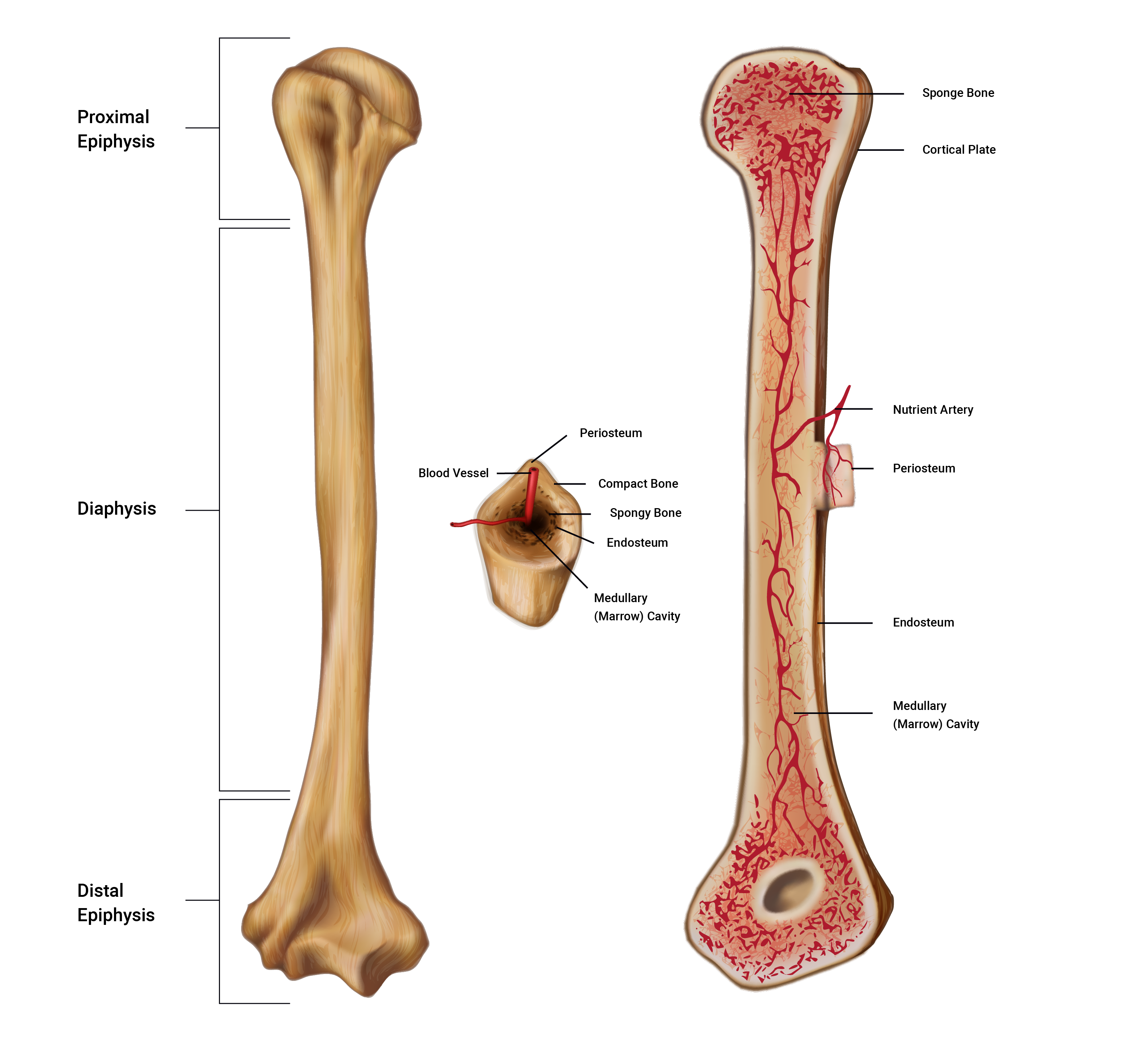
Regions of a long bone explained
The three main regions of a long bone are the proximal and distal epiphyses, the diaphysis and the medullary cavity.
- The epiphyses (e-pif-i-seez): These are found at both the proximal and distal ends of a long bone and named accordingly. The singular is epiphysis. The regions are typically the widest regions of a long bone and are filled with spongy bone (houses red bone marrow) and covered with a shell of compact bone.
- The diaphysis (di- af-y-sis): Known as the 'bone shaft' or 'body', this is the middle section of a long bone. The diaphysis is long, cylindrical, and hollow. The hollow region of the diaphysis is known as the medullary cavity which is where we find yellow bone marrow and many blood vessels ( in adult bones). The hollow design of a long bone enables great strength to be achieved whilst maintaining a long yet lightweight bone which is much easier for our muscles to help move than a heavy dense structure. The diaphysis connects with both the proximal and distal epiphyses.
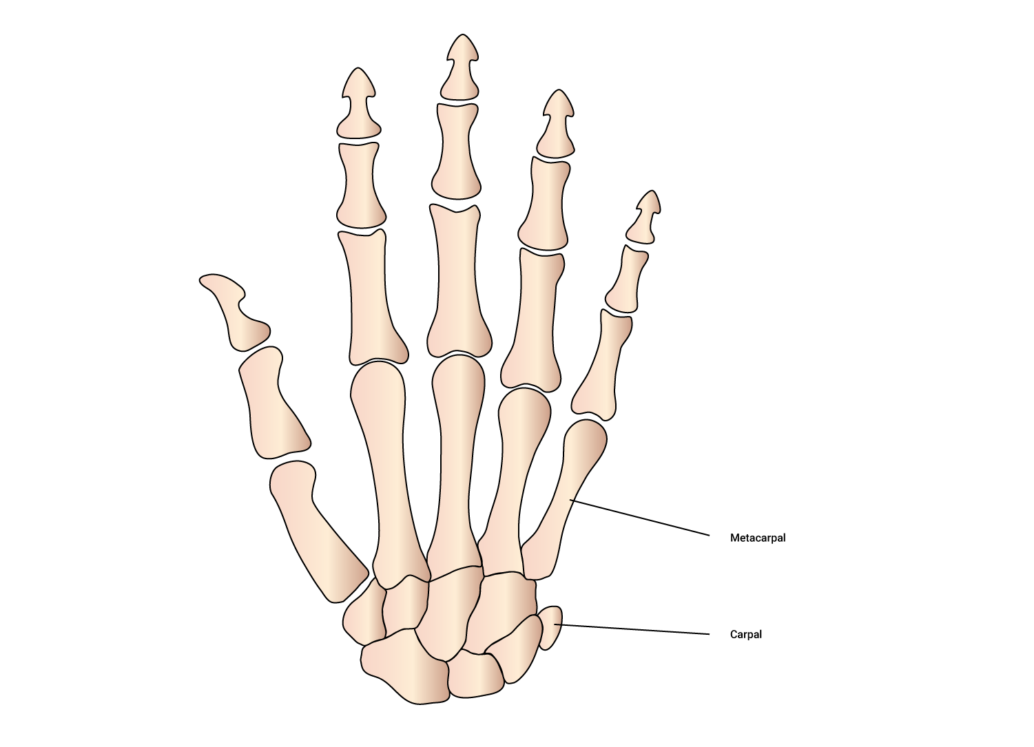
Short bones
These are equal in length and width. They can't move much but can move in most directions a little. Examples of these bones include the:
- carpals (wrist)
- tarsals (feet)
- talus (where the tibia meets the foot).
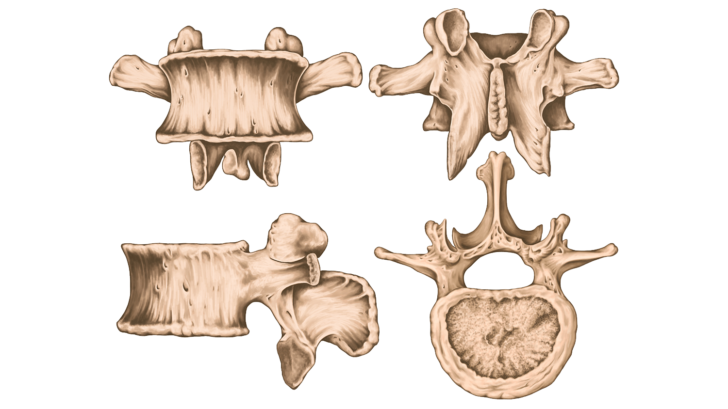
Irregular bones
These vary in shape, they don't really fit into any other category so they have their very own. They often have a complex shape which, thanks to the principle of structure and function, aids their role in protecting internal organs. Examples of these bones include the:
- vertebrae of the spine (protecting the spinal cord)
- pelvic bones (ileum, ischium, and pubic which protect our pelvic organs).
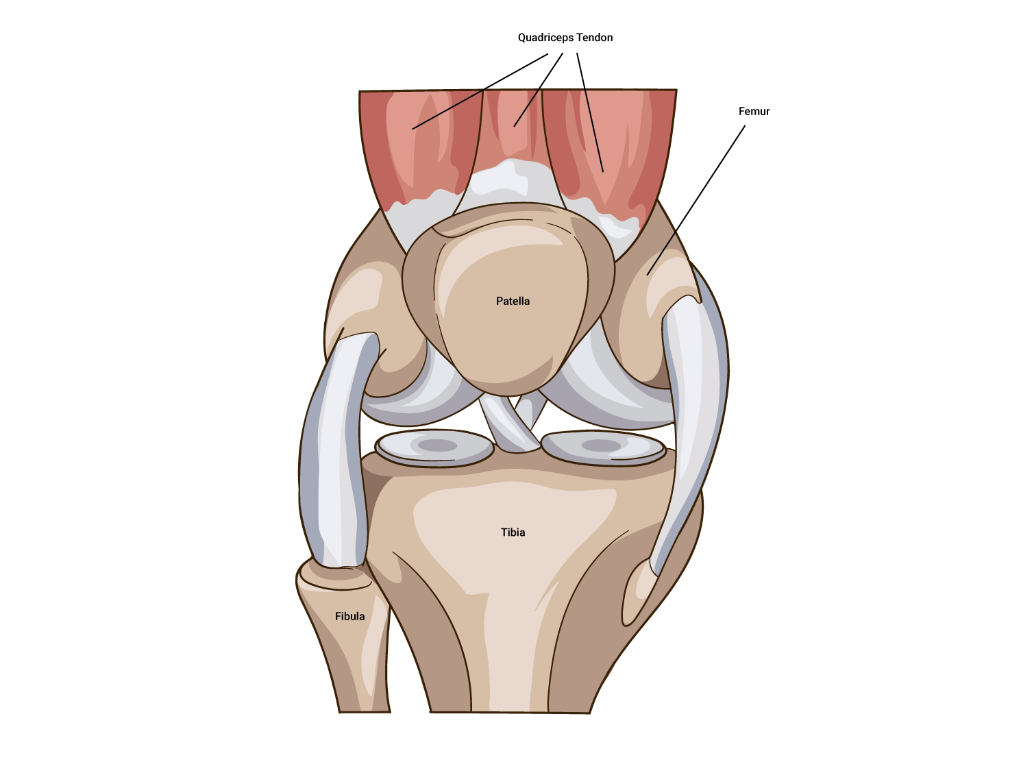
Sesamoid bones
Derived from the Latin sesamum meaning 'sesame seed' due to its typically small size. These bones are often embedded within a muscle or a tendon, for example:
- in the patella (knee cap) which is the most common sesamoid bone
- others can be found in the hand and the foot.
Bone identification
These are the key bones you should know the locations of. Study up on these and add to your personalised terminology and vocabulary reference guide for easy reference.
The adult human skeleton is comprised of 206 named bones. Most of these are paired, that is, they have a 'friend' on the other side of the body, what’s on the left is typically on the right also. The skeletons of infant and children have more bones than in the adult human skeleton. This is because, as our skeletons develop, our bones fuse together (for example, bones of the skull) and therefore the total number of bones decreases.
The bones of the skeleton are anatomically grouped into two different divisions, thus enabling speedy identification and an idea of their role in the skeleton.
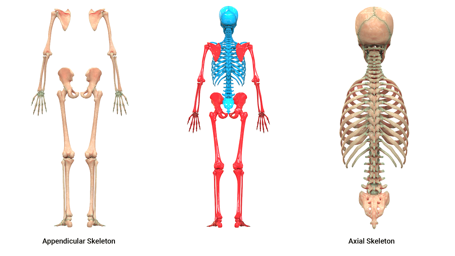
Divisions of the skeleton
- Axial skeleton
- Appendicular skeleton.
Axial skeleton
The axial skeleton is represented in blue in the preceding image. Eighty (80) bones exist within the axial skeleton. The term axial implies axis, therefore we can think of structures down the midline of the body ( the spine is our imaginary midline), therefore the axial skeleton includes bones such as bones of the:
- skull
- face
- auditory bones (ossicles)
- vertebral column
- sternum and ribs.
Appendicular skeleton
The appendicular skeleton is represented in red in the preceding image. These bones comprise of everything that 'hangs off' the axial skeleton, the appendages if you like. The appendicular skeleton consists of 126 bones, which includes the bones of the:
- arms and legs
- pelvis
- hands and feet.
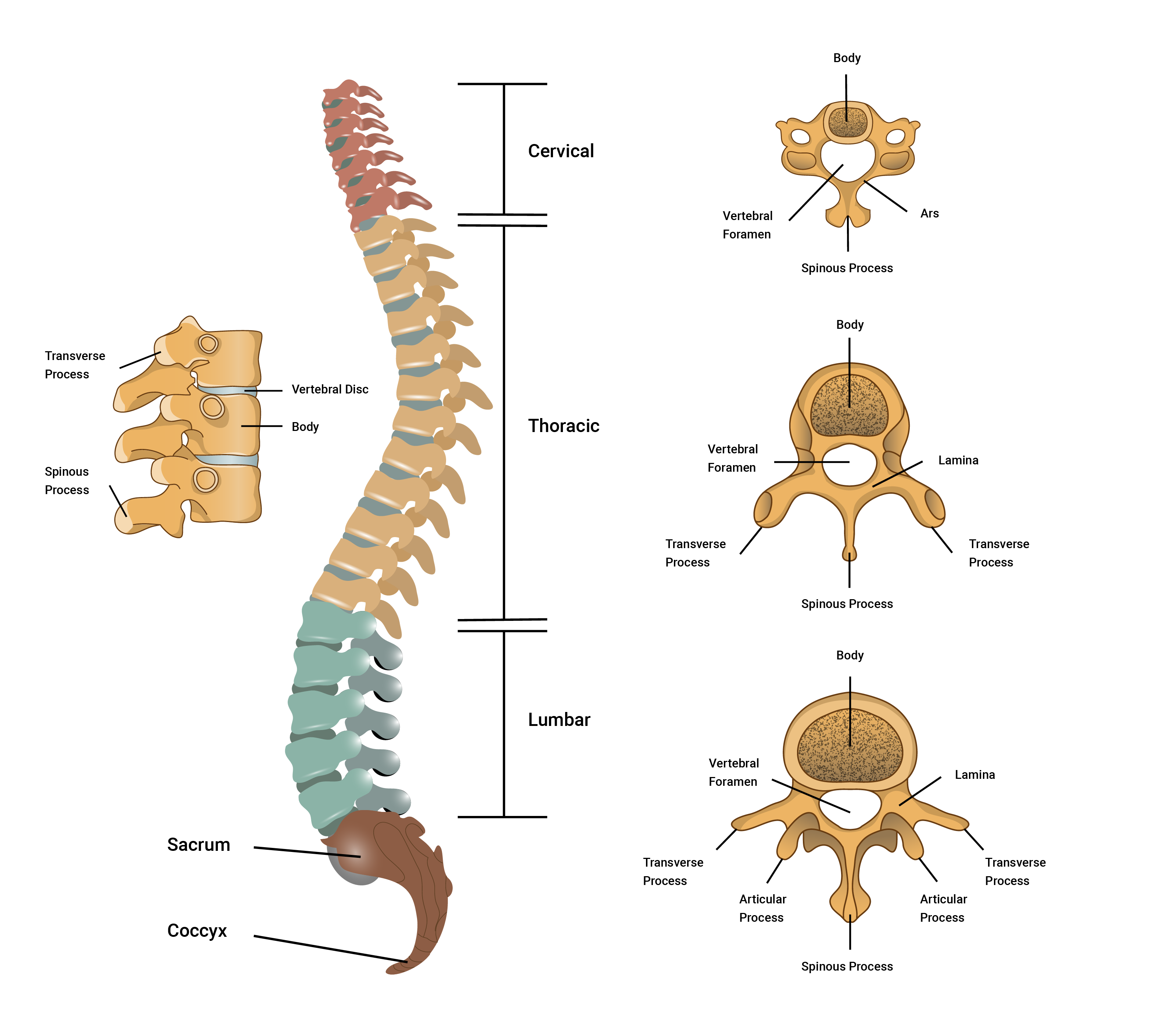
The spine (vertebral column/backbone) makes up approximately two-fifths of your total height.
The spine (vertebral column) is both strong and flexible. It is strong enough to support the head and neck and the body whilst being flexible enough to facilitate movement. It also is the security guard of the spinal cord, a very delicate and fragile structure that runs down the hollow passages of the vertebral column.
The spinal column is composed of 33 independent bones referred to as vertebrae. These vertebrae are divided into five sections where the structure of each vertebra is slightly different due to the functions each performs. The vertebrae are held together by a strong network of ligaments and cushioned by vertebral disks.
Each region of the vertebral column has a specific number of vertebra:
- 7 cervical vertebra in the neck region
- 12 thoracic vertebra in the chest/thorax region
- 5 lumbar vertebrae in the lower back region
- 1 sacrum which consists of five (5) fused sacral vertebrae
- 1 coccyx which consists of four (4) fused coccygeal vertebrae
= 33 irregular independent bones in total.
Remembering these regions, their order, the bones in each region as well as structural changes between the vertebrae of each region is key in anatomy and physiology. This knowledge may not be called upon every day but it underpins the rest of your anatomy, physiology and exercise-based knowledge. Therefore, it is important to become comfortable with these:
A handy mnemonic to remember the regions and the number of vertebrae in each:
You have Cereal at 7, Tea (cuppa) at 12, Lunch at 5 (ok, it’s a late lunch) and then a Cuddle on the Sofa at 11 (11 = 1 + 1 of the sacrum and coccyx)
Vertebral joints
Each region of the spine has varying degrees of movement due to structural differences and the presence of ribs in the thoracic region.
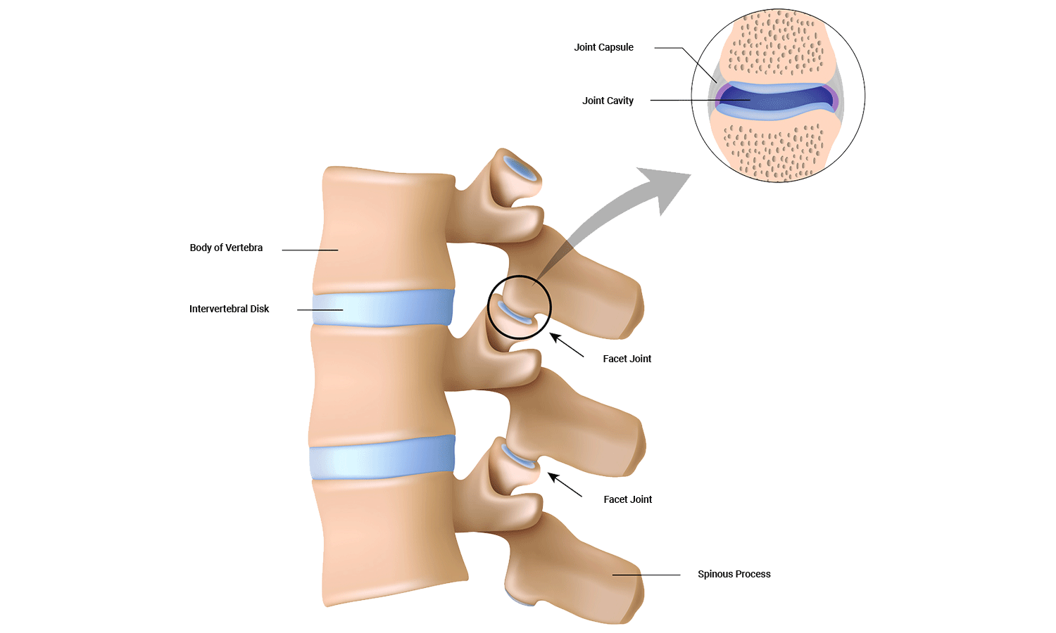
Facet joints
Located between the superior facet of one vertebra and the inferior facet of the vertebrae (refer to preceding image). Approximately 16% of the load of the body is absorbed by these joints. If they are poorly aligned, or under chronic extra stress, these can be the location of common back pain.
Intervertebral joints
Inter-vertebral joint ( inter~ in between, vetebral~vertebra): This joint is found between the vertebral bodies of the vertebrae. Eighty-four per cent (84%) of the load of the body should be absorbed here (if well aligned).
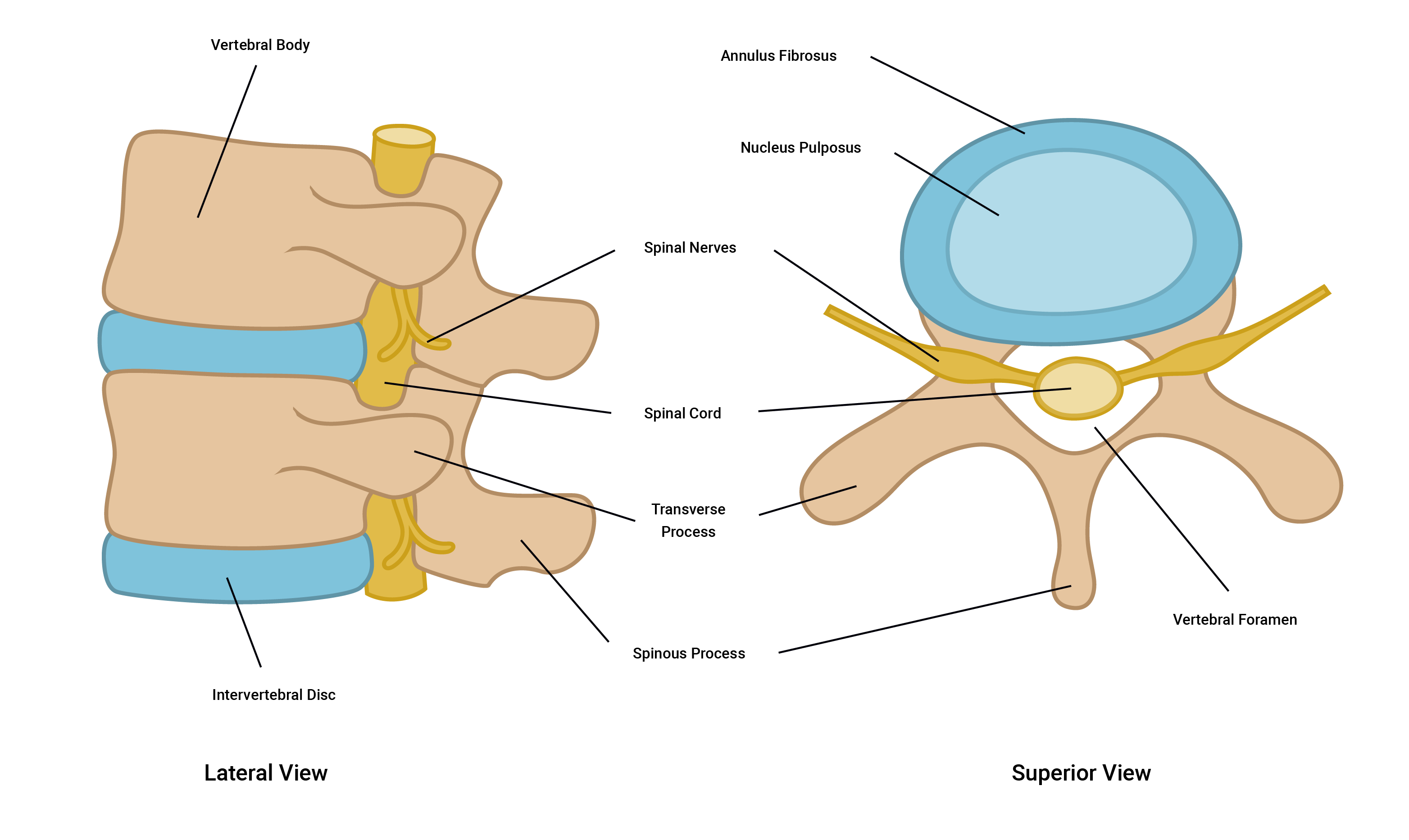
Intervertebral discs
There are three joints between each vertebra; two facet joints and one inter-vertebral joint. Each inter-vertebral joint is separated by an intervertebral disc. Together as a team, the intervertebral discs and the vertebra form the vertebral column.
The intervertebral discs are comprised of two main components:
- The annulus fibrosus: The annulus fibrosus are tough fibro-cartilage rings. These are very strong under compression, but susceptible to torsional (twisting) stress.
- The nucleus pulposus: The nucleus pulposus is mainly made up of water (90%) and provides shock absorption like water-filled cushions.
Fun fact
Disc height changes over the course of a day due to axial loading. This results in us being taller in the morning than we are at night!
Enjoy this short yet stunning 3D video of the human skeleton looking at the different regions of the spine.
Use it or lose it!
If little stress is placed on bones, bones will demineralise. This can happen for a variety of reasons including:
- sedentary lifestyle
- low impact endurance exercise
- becoming bed-ridden.
Nutritional deficiency can also contribute to this.
Bone wasting conditions
Bone growth is dependent on sex hormones. As the levels of these hormones reduce with age, bone mineralisation decreases and bone atrophy (wasting) can start to occur.
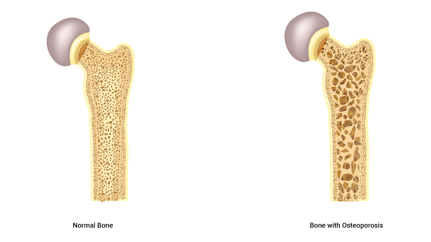
All of us will eventually suffer from osteopenia which is a natural de-mineralisation of bones as we get older.
If this gets to the point where daily living is compromised it is known as osteoporosis.
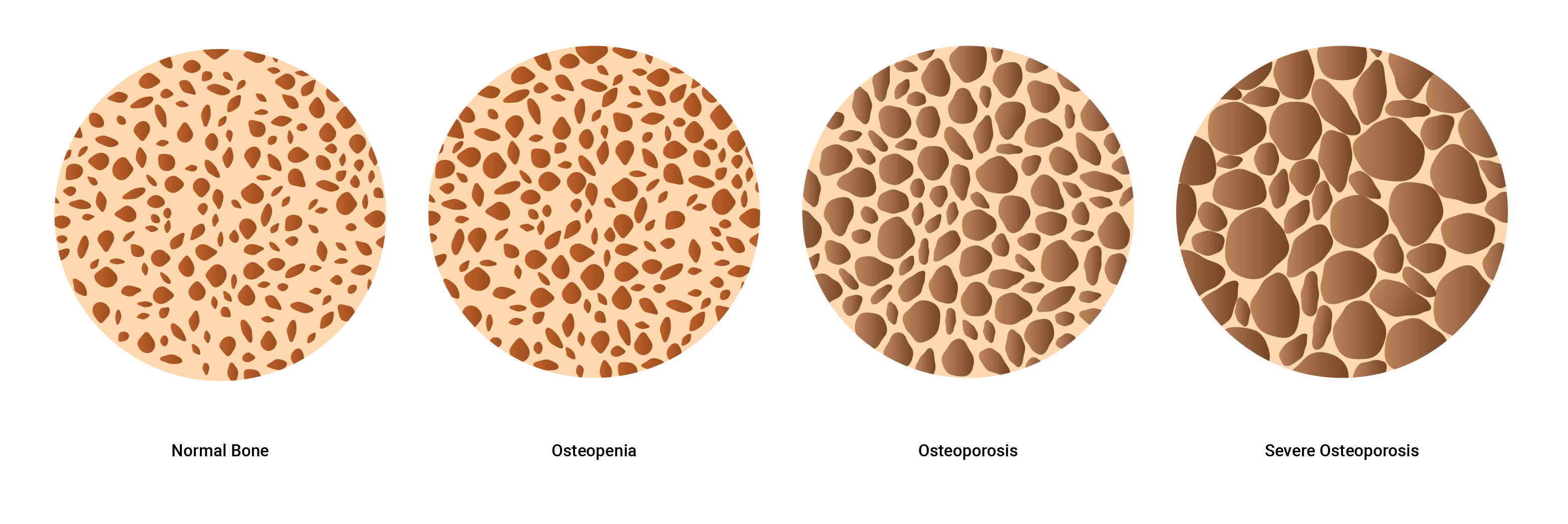
The term 'bone mineral density' refers to the amount of bone mineral (mainly calcium) present in a bone. Bones that have high mineral density are stronger, can withstand greater forces, and are less likely to fracture. Bones with low mineral density are weaker and more prone to fracture under stress.
Factors contributing to bone density
Three factors are critical in developing bone mineral density:
- Sex hormones: Testosterone and estrogen hormones (among others) stimulate bone deposits.
- Nutrition: Adequate food intake including regular consumption of calcium.
- Loaded exercise: This refers to exercise that places repetitive stress on bones to encourage bones to adapt (strengthen).

Research has clearly shown a direct relationship between exercise and bone health. But not all exercise affects bone the same when it comes to building strong, healthy bones or preventing osteoporosis.
Loaded exercise is the key to maintaining bone health (density).
The term 'loaded' refers to an activity that places at least bodyweight on bones, such as running, running-based sports, skipping, and weight lifting. When placed under stress (mechanical stress) bone tissue becomes stronger through the increased rate of bone production, which occurs quicker than the bone reabsorption (breakdown) phase of the bone remodelling process. Therefore activities such as jumping and running stimulate the process of bone remodelling more effectively and efficiently than activities such as low weight-bearing activities (for example, cycling and water-based activities) because these do not elicit the same response in terms of bone density.
In order to stimulate bone growth, the exercise must elicit stress on the bone. The other key factor in bone health is nutrition. You must provide your body with enough essential minerals to support bone growth and maintenance.
People who do regular loaded physical activity produce denser and more mineralised bones when compared with sedentary individuals.
Occupations and sports that are predominantly one arm dominant (for example, tennis players baseball pitchers, or cricket bowlers) result in more mineralised bones on the dominant side.
Three key factors to consider when selecting exercises to enhance bone mineral density

Three characteristics of exercise that have the largest impact on increased bone density are:
- strain magnitude of the exercise
- strain rate of the exercise
- strain frequency of the exercise
Strain magnitude
The amount of stress placed on the bones.
This is higher in exercise such as gymnastics and weightlifting where the force or impact of the exercise is greatest
Strain rate
This is the velocity over which the strain is placed on the bones.
This is higher in exercise such as jumping or plyometrics where the rate at which the impact is felt is high.
Strain frequency
This is how often strain is placed on bones during the course of an activity.
This is higher in exercise such as running, where the impact on the bones occurs frequently during the exercise session.
Order of importance
Strain magnitude is the most important of the three factors for bone density. Velocity is the next most important factor followed by frequency.
- Strain magnitude
- Strain rate (velocity)
- Strain frequency.
Cross-training
Athletes involved in sports that have low strain magnitude (such as water-based sports and cycling) would benefit from a form of cross-training that is higher in strain magnitude. For example, a water polo player may benefit from doing some weight training to ensure their bone density is maintained.
Cross-training is a form of training which typically incorporates multiple training systems through a variety of exercises to develop and enhance overall performance.
It is important to have the correct balance of these when designing training programs. Consider a thorough pre-screening session when you work with both new and existing clients.
It’s important in developing a safe and effective exercise program design, to take the time to sit down with your client, take them through a pre-screening questionnaire and consider providing an example (discussed in a later topic). Break the ice with the client to gain a thorough (but not intrusive) medical history to ensure you are aware of any potential risks you can avoid, by selecting the correct exercise and exercise frequency. This comes under the mindset of program design and pre-screening.
In this topic, we focused on the structure and function of the skeletal system. You learnt about:
- the different functions and shapes of bones
- how to identify types and location of bones
- the structure and function of the spine
- effect of exercise on bone density including osteoporosis and exercise considerations
- body cavities, planes and sections
- movement types and directional terms.